Porokeratotic eccrine ostial and dermal duct nevus
– Porokeratotic eccrine duct and hair follicle nevus
– Porokeratotic adnexal ostial nevus
Clinical Issues
• Congenital or develop in childhood; may present in adulthood
Microscopic
• Eccrine angiomatous hamartoma
Eccrine glands normal (occasionally enlarged) or increased in number
Endothelial cell-lined, thin-walled vascular spaces intimately admixed with eccrine glands
• Coccygeal polypoid eccrine nevus
Polypoid shape; increased number of eccrine glands
• Porokeratotic eccrine ostial and dermal duct nevus
Cornoid lamellae (tiers of parakeratosis over diminished granular layer) above acrosyringeal ducts &/or hair follicles
• Eccrine nevus
Increased number or size of eccrine glands
No increase in vessels; not polypoid
Top Differential Diagnoses
• Eccrine angiomatous hamartoma
Eccrine nevus
• Eccrine nevus
Normal eccrine glands
• Coccygeal polypoid eccrine nevus
Skin tag/acrochordon or soft fibroma
• Porokeratotic eccrine ostial and dermal duct nevus
Porokeratosis
Inflammatory linear verrucous epidermal nevus
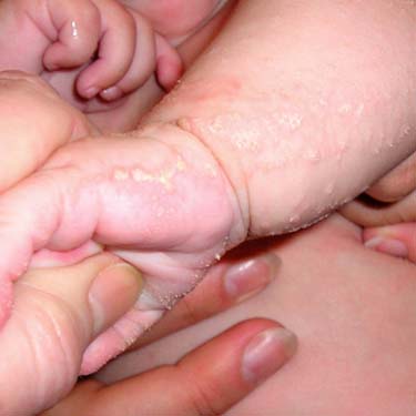
Clinical Photograph of Porokeratotic Eccrine Ostial and Dermal Duct Nevus This is a porokeratotic eccrine ostial and dermal duct nevus with light pink, hyperkeratotic streaks following Blaschko lines. There are focal spines. (Courtesy J. McNiff, MD, and R. Antaya, MD.)
Porokeratotic Eccrine Ostial and Dermal Duct Nevus Biopsy of porokeratotic eccrine ostial and dermal duct nevus shows tiers of parakeratosis (cornoid lamellae) overlying acrosyringeal ostia. The inset shows another example with a cornoid lamella. (Courtesy J. McNiff, MD.)
Eccrine Angiomatous Hamartoma In eccrine angiomatous hamartoma, the epidermis is often normal; this example has an overlying verrucous hemangioma . In the lower dermis, eccrine glands are intimately admixed with fat and vessels (as seen in the inset). (Courtesy of A. Galan, MD.)
Coccygeal Polypoid Eccrine Nevus Coccygeal polypoid eccrine nevus is polypoid; increased numbers of eccrine glands are in the dermis (highlighted in the inset).
TERMINOLOGY
Synonyms
• Eccrine angiomatous hamartoma
Sweating angiomatous hamartoma
Sudoriparous angioma
Functioning sudoriparous angiomatous hamartoma
• Porokeratotic eccrine ostial and dermal duct nevus
Porokeratotic eccrine duct and hair follicle nevus
Only gold members can continue reading. Log In or Register to continue


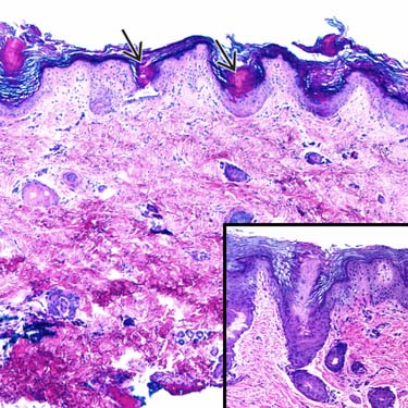
 overlying acrosyringeal ostia. The inset shows another example with a cornoid lamella. (Courtesy J. McNiff, MD.)
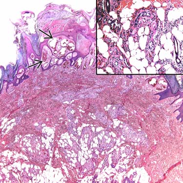
 . In the lower dermis, eccrine glands are intimately admixed with fat and vessels (as seen in the inset). (Courtesy of A. Galan, MD.)
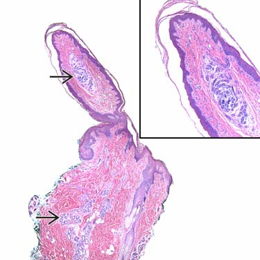
 are in the dermis (highlighted in the inset).


