QUESTION 23.1
A. Chondrodermatitis nodularis chronica helicis
B. Gout
C. Ochronosis
D. Malakoplakia
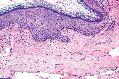
E. Spectacle frame acanthoma
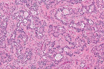
2. A mass is removed from the middle ear of a 48-year-old woman. This photomicrograph shows its histopathologic features. Which of the following is the most likely diagnosis?
QUESTION 23.2
A. Adenoma of the middle ear
B. Cholesteatoma
C. Cholesterol granuloma
D. Dermoid cyst
E. Squamous cell carcinoma
3. A 47-year-old man presents with tinnitus and painful swelling of both ears. He reports past episodes of pain and swelling of the nose as well. He also complains of recurrent debilitating joint pains. On physical examination, “cauliflower” deformities of both ears and a “saddle” nose are noticed. The patient is found to have bilateral sensorineural hearing loss. A biopsy of the ear shows the histopathologic changes depicted in this photo. Which of the following is the most likely pathogenetic mechanism of this condition?
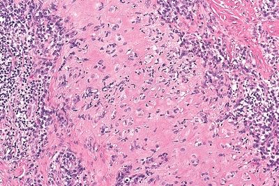
QUESTION 23.3
A. Altered purine metabolism
B. Autoimmune
C. Genetic
D. Infectious
E. Neoplastic
4. A total stapedectomy is performed for treatment of otosclerosis in a 45-year-old male. Which of the following histologic changes is most likely to be present?
A. Extensive osteolysis of the footplate.
B. New woven bone formation in the crura.
C. New woven bone formation in the footplate.
D. Spongiosis and formation of large marrow cavities.
E. No changes: Pathologic changes are limited to otic capsule.
5. A 40-year-old woman undergoes excision of slow-growing, skin-covered polypoid nodule in the external auditory canal. Its morphologic appearance is shown in this picture. Which of the following is this tumor?
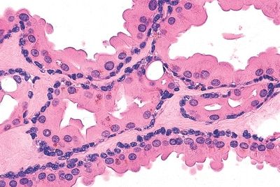
QUESTION 23.5
A. Adenoid cystic carcinoma
B. Basal cell carcinoma
C. Ceruminous gland adenocarcinoma
D. Ceruminous gland adenoma
E. Mucoepidermoid carcinoma
F. Squamous cell carcinoma
G. Syringocystadenoma papilliferum
6. A 30-year-old woman presents with a pruritic subcutaneous nodule in the left auricle. There is no regional lymphadenopathy. Local surgical excision is performed. The histologic appearance of the lesion is shown in this photomicrograph. Which of the following is the most likely diagnosis?
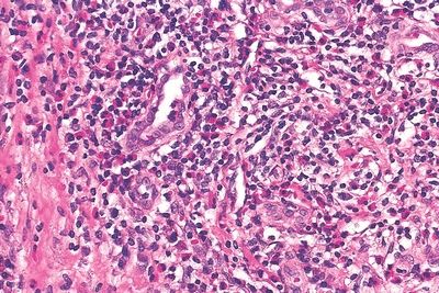
QUESTION 23.6
A. Epithelioid angiosarcoma
B. Epithelioid hemangioendothelioma
C. Epithelioid hemangioma
D. Lobular capillary hemangioma
7. A small tumor is removed from the middle ear of a 40-year-old man suffering from conductive hearing loss. This picture demonstrates its microscopic features. Which of the following is the most likely diagnosis?
QUESTION 23.7
Stay updated, free articles. Join our Telegram channel

Full access? Get Clinical Tree


