• Histology usually improves with cessation of offending drug
Microscopic
• Noncaseating granulomas
Often associated with lymphocytes, plasma cells, and (most notably) eosinophils
– Presence of granulomas, ± eosinophils, does not prove drug-related etiology, however
Hepatocyte reactive changes, apoptotic hepatocytes, cholestasis, cytoplasmic ballooning/feathery degeneration may be present
– Combination of microgranulomas and hepatocyte injury is very suggestive of granulomatous drug reaction
Diagnostic Checklist
• Careful drug history and temporal correlation between drug administration and liver disease are essential
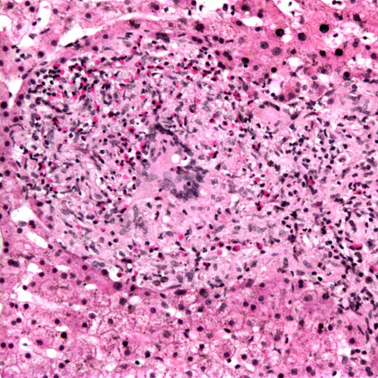
Epithelioid Granuloma With Eosinophils This portal tract contains an epithelioid granuloma with numerous associated eosinophils, as well as a central giant cell. The patient’s granulomatous hepatitis was due to drinking Echinacea (coneflower) tea.
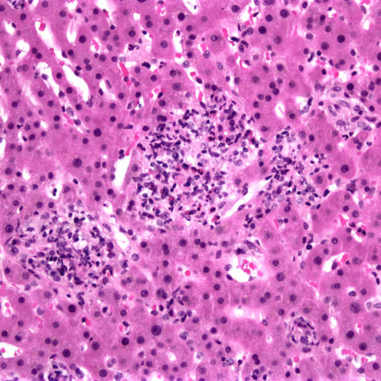
Microgranuloma Microgranulomas, shown here with admixed lymphocytes and eosinophils, are often seen in drug reactions. This patient had a reaction to propylthiouracil. Microgranulomas are often accompanied by hepatocyte injury.
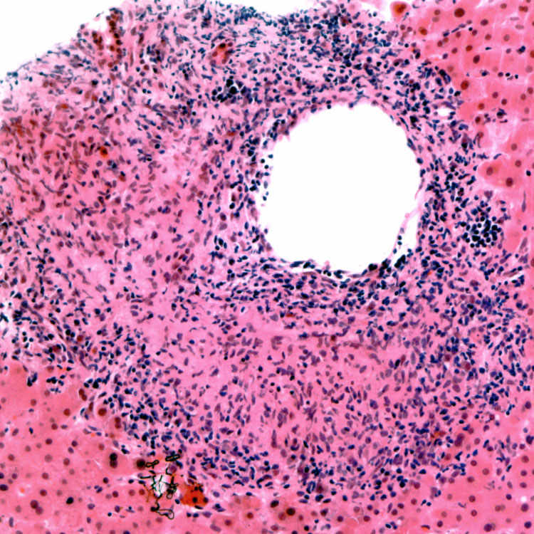
Granulomatous Vasculitis Granulomatous vasculitis surrounding a central vein is seen in this case of granulomatous hepatitis due to allopurinol. (Courtesy J. Misdraji, MD.)
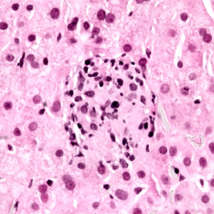
Small Portal Granuloma A small portal epithelioid granuloma with associated lymphocytes is seen in this case of Tegretol-related granulomatous hepatitis.
TERMINOLOGY
Definitions
• Granulomatous inflammation caused by drug or toxin
Important mechanism of drug-related hepatotoxicity
Drugs reportedly responsible for up to 30% of hepatic granulomas
Many implicated drugs, including over-the-counter and herbal preparations
ETIOLOGY/PATHOGENESIS
Probable Hypersensitivity Reaction
• Common offenders
Antimicrobials
Only gold members can continue reading. Log In or Register to continue