Disorders of the Eyes, Ears and Other Sensory Organs
Learning Objectives
After studying this chapter, the student is expected to:
1. Define sensory receptors and classify them by location and stimuli.
3. Describe common infections in the eye and their possible effects on vision.
4. Explain how intraocular pressure may become elevated and how it may affect vision.
6. Describe how the retina may become detached and the possible effects on vision.
7. Describe the types of hearing loss with an example of each.
8. Describe otitis media and its cause, pathophysiology, and signs.
9. Describe the pathophysiology and signs of otosclerosis and of Ménière’s syndrome.
Key Terms
diplopia
exteroceptors
ototoxic
photophobia
proprioceptors
ptosis
refraction
tinnitus
trachoma
visceroceptors
visual acuity
Sensory Receptors
Sensory receptors/sense organs are classified into two categories: general senses and special senses. Special senses include the eye and the ear.
Sensory receptors can be classified by their location
• Visceroceptors (interoceptors)
• Located internally and provide information about the environment around the viscera.
Sensory receptors are classified by their stimuli:
• Stimulated by a mechanical force: touch, pressure, equilibrium, hearing
• Activated by a change in chemical concentration
• Stimulated by a change in temperature
• Respond to any tissue damage, the sensation produced is pain
The Eye
Review of Structure and Function
Visual information is received from light rays that pass through the transparent cornea and then through the lens, which focuses the image on the receptor cells of the retina, the rods and cones. These visual stimuli are conducted by the optic nerves to the occipital lobe of the brain for interpretation and processing before they are sent to other appropriate areas of the brain.
Protection for the Eye
The eye is well protected in the bony orbit of the skull. The eyelids (palpebrae) and eyelashes deflect foreign material in the air away from the eyes and protect the eye from excessive sunlight and drying. The levator palpebrae superior, the muscle of the upper eyelid, is controlled by the oculomotor nerve (cranial nerve III). The eyelids are lined with a thin mucous membrane, the conjunctiva, which continues over the sclera of the eye.
The continual secretion of tears washes away particles, microbes, and irritating substances. This watery secretion contains lysozyme, an antibacterial enzyme. The tears form in the lacrimal gland located on the superior lateral area of the orbit. They flow across the eye and drain into the lacrimal canals in the medial corner of the eye and then into the nasal cavity through the nasolacrimal duct. The tears keep the external tissues of the eye moist and healthy.
The Eyeball
Six skeletal muscles (extrinsic muscles) control the movement of the eyeball in the bony orbit. They originate on the orbit and insert onto the sclerae on the outside of the eyeball. There are four straight (rectus) muscles and two angled (oblique) muscles, which are coordinated to move and rotate the eye and are under the control of cranial nerves III, IV, and VI.
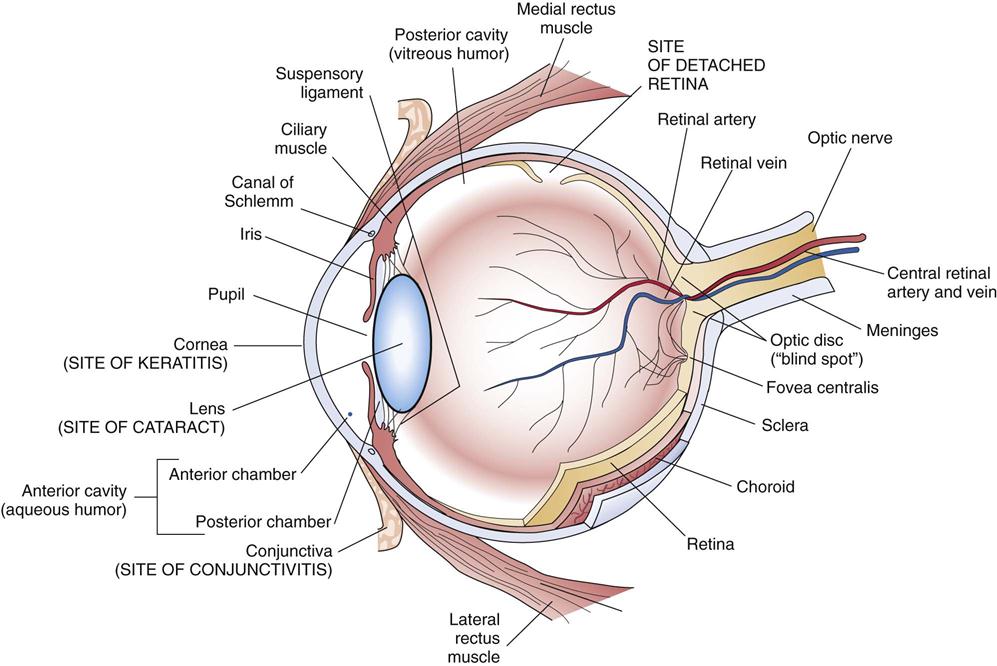
The eyeball consists of a spherical three-layered wall filled with fluid (Fig. 15-1):
In the anterior part of the eye, the choroid develops into the ciliary body and iris. The ciliary body consists of the ciliary muscle, which controls the shape of the lens to focus the image of near and distant objects accurately and clearly on the retina, and the ciliary processes, which secrete aqueous humor, the fluid in the anterior cavity of the eye.

The iris is a circular structure surrounding the pupil, an opening through which light rays pass into the interior of the eye. The iris is pigmented and gives the eye its distinctive brown or blue tone. The iris contains two muscles that control the size of the pupil. The circular, or sphincter, muscle contracts in response to parasympathetic stimuli or excessive light on the retina, resulting in a constricted or small pupil. Parasympathetic nervous system fibers reach the iris from cranial nerve III. Under sympathetic nervous system control, the radial muscles of the iris, when contracted, cause the pupil to dilate or open. This function is easier to remember if it is associated with the stress (GAS, see Chapter 26) or fight-or-flight response, in which sympathetic stimulation leads to pupil dilation and improved vision, especially in dim light.
The lens is a transparent biconvex structure made up of an elastic capsule surrounding an orderly alignment of fibers. There are no blood vessels or nerves to interfere with the transparency, nor are there organelles in the cells. However the lack of organelles limits repair in these cells. Nutrients are provided from the aqueous humor. Together with the cornea, the lens provides refractive power for the light entering the eye. Current interest relates to the mechanism by which these cells have lost the organelles and become transparent.
The suspensory ligament connects the lens to the ciliary body. The shape of the lens is altered as the contraction of the ciliary muscle alters tension on the suspensory ligament. This adjustment in lens curvature, or accommodation, bends the light rays (refraction) entering the eye sufficiently to focus a sharp image on the retina.
The innermost layers of the retina consist of the rods and cones, which are light-sensitive photoreceptor nerve cells. Rods are specialized for dim light (for night vision), and cones are color sensitive. The light energy is absorbed by the rods and cones and converted into electrical energy in the neurons. Three types of cones, red, green, and blue, determine color perception. Color blindness is common in males and results from a deficit of one type of cone owing to an abnormal gene on the X chromosome (sex-linked recessive gene) (see Chapter 21).
Fluids in the Eye
The eye is divided into two cavities by the lens and ciliary body:
The anterior cavity is filled with aqueous humor, which is continuously secreted by the ciliary processes into the posterior chamber. It flows through the pupil into the anterior chamber and drains into the reticular network and canal of Schlemm (see Fig. 15-4). This canal encircles the eye at the junction of the cornea and iris and returns the fluid to the blood. To maintain normal intraocular pressure inside the eye (normal range 12 to 20 mmHg, average 15 mmHg), the amount of aqueous humor formed should equal the amount reabsorbed. Normal pressure maintains the shape of the eye. The aqueous humor supplies nutrients to the lens and cornea, which lack blood vessels.
The Visual Pathway
To review the physiology of vision, light rays from an object pass through the cornea, where they are refracted, and then through the aqueous humor and pupil. The curvature of the lens is adjusted to refract the light rays so that they converge on the retina, providing a sharp image of the object. The light continues through the transparent aqueous humor to the retina, where the photoreceptor neurons, the rods and cones, are stimulated. The light energy is converted into an electrical stimulus, which is transmitted by the optic nerve to the occipital lobe of the brain, where the image is identified and integrated with other information. The double image projected from different angles by the two eyes provides a wider visual field, and the central overlap of visual fields provides depth perception.
The nerve impulses from the ganglion cells of the retina converge in the fibers of the optic nerve (cranial nerve II), which leaves the eye at the optic disc in the posterior portion of the eye. The central retinal artery and vein, which supply the retina and other structures, also pass through the optic disc. There are no rods or cones at the optic disc, forming the “blind spot.”
Because the optic nerve is essentially a projection of brain tissue surrounded by cerebrospinal fluid and meninges, it reflects pressure in the brain (see Chapter 14). Assessment of the eye can often provide useful information about other problems in the body, such as hypertension or vascular changes as a result of diabetes.
The optic nerves carry visual stimuli to the occipital lobes of the brain (see Fig. 14-1). At the optic chiasm, half of the fibers from each optic nerve cross to pass to the occipital lobe in the opposite hemisphere (see Fig. 14-7). Therefore the left occipital lobe receives images from the right visual field. Damage to the left occipital lobe results in loss of the right visual field.
Diagnostic Tests
Basic tests include the following:
• Visual field tests are used to check central and peripheral vision.
• Tonometry assesses intraocular pressure by checking the resistance of the cornea.
• An ophthalmoscope can be used to examine the interior structures.
• Gonioscopy measures the angle of the anterior chamber.
Many other sophisticated tests are available for specific disorders. Neurologic damage to the visual pathway is covered in Chapter 14. Retinopathies are discussed in Chapter 12 under hypertension, and in Chapter 16 under diabetes mellitus.
Structural Defects
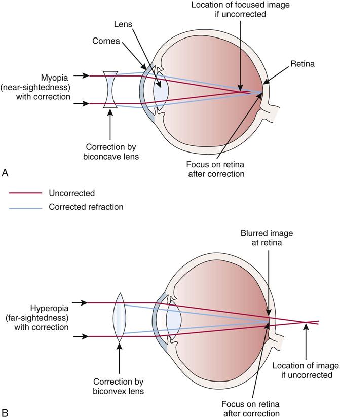
Structural defects interfere with the focusing of a clear image on the retina:
• Myopia, nearsightedness, occurs when the image is focused in front of the lens, perhaps because the eyeball is too long (Fig. 15-2).
• Astigmatism develops from an irregular curvature in the cornea or lens.

Infections and Trauma
A stye (hordeolum) is an infection involving a hair follicle on the eyelid, usually by staphylococci. A swollen, red mass forms on the eyelid, which is uncomfortable when blinking.
Conjunctivitis
Conjunctivitis is a superficial inflammation or infection involving the conjunctiva lining the eyelids and covering the sclera. Allergens, bacteria, viruses, or irritating chemicals in the air are a frequent cause of inflammation, resulting in redness, itching, and excessive tearing with a watery discharge.
Organisms such as Staphylococcus aureus cause the highly contagious “pinkeye,” which occurs frequently in children. The sclera of the eye and eyelid appears red, and there is a purulent discharge. Pinkeye is spread by the fingers or contaminated towels. Antibiotic treatment is required to reduce contagion and prevent damage to the cornea.
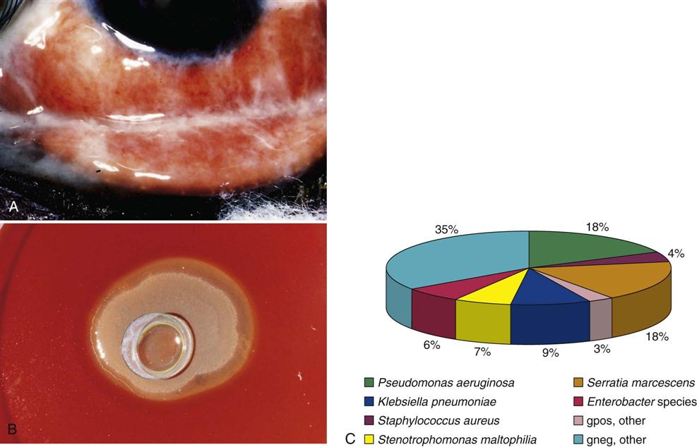
Contact lenses are a frequent source of infection in the eye, both conjunctivitis and keratitis (Fig. 15-3B,C). Other sources are contaminated medication or makeup. Medications or eye makeup should not be used past the expiry date or instructions. The infectious agent could be one of a variety of microorganisms.
Stay updated, free articles. Join our Telegram channel

Full access? Get Clinical Tree


