QUESTION 5.7
A. Fibrosarcoma
B. Keloid scar
C. Myxoid liposarcoma
D. Myxoid malignant fibrous histiocytoma
E. Nodular fasciitis
8. The type of lipoma most likely to mimic a sarcoma is:
A. Angiolipoma
B. Fibrolipoma
C. Myxolipoma
D. Pleomorphic lipoma
E. Spindle cell lipoma
9. This picture shows the characteristic cellular composition of a lipoma type usually found on the back of the neck of elderly men. Which one is it?
QUESTION 5.9
A. Angiolipoma
B. Fibrolipoma
C. Myxolipoma
D. Pleomorphic lipoma
E. Spindle cell lipoma
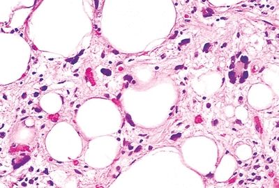
10. A review of slides of a tumor excised from the upper extremity of a 2-year-old child and previously diagnosed as myxoid liposarcoma reveals a neoplastic lesion composed of variably differentiating lipoblasts, as seen in this photomicrograph. The architecture is distinctly lobular, with a myxoid background containing a plexiform vascular network. Which of the following is the most likely diagnosis?
QUESTION 5.10
A. Angiolipoma
B. Hibernoma
C. Lipoblastoma
D. Myxoma
E. Well-differentiated liposarcoma
11. The fetal form of rhabdomyoma may develop in the vulvovaginal region of middle-aged women. Which of the following is the most common location in children?
A. Extremities
B. Head and neck
C. Mediastinum
D. Perineal region
E. Retroperitoneum
12. Which of the following vascular lesions is thought to arise as a florid form of organized and recanalized thrombus?
A. Capillary hemangioma
B. Intravascular papillary endothelial hyperplasia
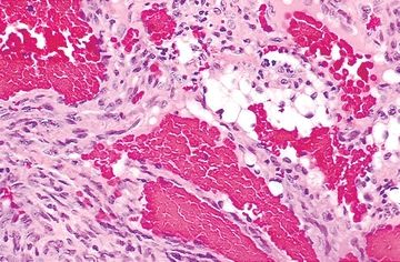
C. Malignant endovascular papillary angioendothelioma
D. Spindle cell hemangioendothelioma
E. Symplastic hemangioma
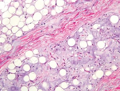
13. A 50-year-old woman undergoes excision of a slowly growing, poorly circumscribed mass in the thigh. The tumor consists of abundant myxoid matrix containing scanty cells and rare vessels, as shown in this picture. Which of the following is the most likely diagnosis?
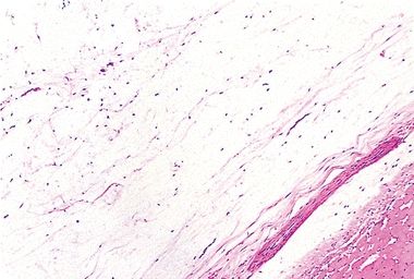
QUESTION 5.13
A. Intramuscular myxoma
B. Myxofibrosarcoma
C. Myxoid chondrosarcoma
D. Myxoid liposarcoma
E. Neurothekeoma
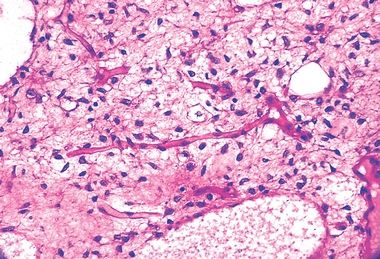
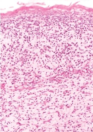
14. A poorly circumscribed nodular mass is removed from the right subscapular region of a 65-year-old man. The lesion lies beneath the latissimus dorsi muscle. Histologically, it consists of thick bundles of collagen intermixed with eosinophilic cylindrical structures, which on elastin stain appear as in this picture. Which of the following is the most likely diagnosis?
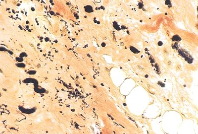
QUESTION 5.14
A. Elastofibroma
B. Fibroma of the tendon sheath
C. Fibromatosis
D. Keloid scar
E. Nodular fasciitis
15. A small subcutaneous nodule removed from the wrist of a 13-year-old male consists of spindly, fibroblast-like cells with spotty calcifications. The cells surrounding calcific foci resemble chondrocytes. Cytologic atypia and mitotic activity are absent. Peripheral infiltration of muscle is seen. Which of the following is the most likely diagnosis?
A. Calcifying aponeurotic fibroma
B. Fibroma of the tendon sheath
C. Fibromatosis
D. Rheumatoid nodule
E. Schwannoma
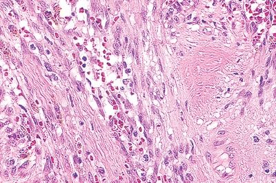
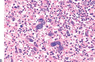
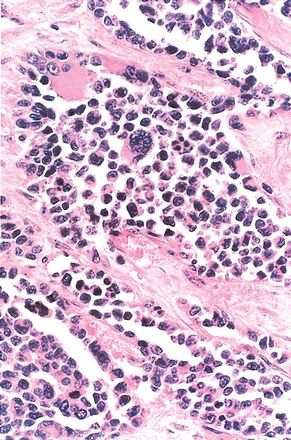
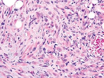
16. Histologically, a retroperitoneal mass from a 40-year-old man consists of fascicles of spindly cells in a sea of lymphocytes, foamy macrophages, plasma cells, and eosinophils, as shown in this picture. The spindly cells are immunoreactive with the p80 antibody to anaplastic lymphoma kinase (ALK) protein. These features are most consistent with:
QUESTION 5.16
A. Desmoid fibromatosis
B. Inflammatory myofibroblastic tumor
C. Nodular fasciitis
D. Palisaded myofibroblastoma
E. Solitary fibrous tumor
17. Ultrastructural and immunohistochemical studies indicate that the principal cell constituents of fibromatosis are:
A. Fibroblasts
B. Histiocytes
C. Myofibroblasts
D. Schwann cells
E. Smooth muscle cells
18. Immunohistochemically, solitary fibrous tumors are typically:
A. Positive for CD34 and negative for SMA
B. Positive for SMA and negative for CD34
C. Positive for β-catenin and negative for STAT6
D. Negative for both BCL-2 and CD99
19. The myofibroblastic lesion depicted in this photomicrograph is exclusively found in the:
QUESTION 5.19
A. Inguinal lymph nodes
B. Mediastinum
C. Pleura
D. Retroperitoneal space
E. Subcutaneous tissue
20. One of the most characteristic morphologic features of fibrosarcomas is:
A. Herringbone pattern
B. Multinucleated giant cells
C. Palisading of cells
D. Serpentine nuclei
E. Storiform pattern
21. Which of the following types of fibrosarcomas arises in the hands and feet and is associated with the characteristic histologic picture shown here?
QUESTION 5.21
A. Low-grade fibromyxoid sarcoma
B. Myofibrosarcoma
C. Myxofibrosarcoma
D. Myxoinflammatory fibroblastic sarcoma
E. Sclerosing epithelioid fibrosarcoma
22. Which of the following microscopic patterns is most characteristic of dermatofibrosarcoma protuberans (DFSP)?
A. Cartwheel/storiform pattern with no significant atypia
B. Long spindle cell fascicles in a herringbone pattern
C. Parallel strands of keloidlike collagen
D. Spindle cell tumor with giant rosettes
E. Zonal architecture
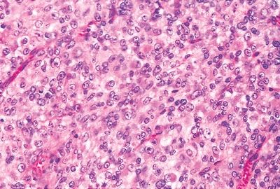
23. A rapidly growing soft tissue tumor is excised from the thigh of a 56-year-old man. It is attached to the muscular fascia, appears well circumscribed but not encapsulated, and measures 14 cm in greatest diameter. On low power, the neoplasm exhibits a vague storiform arrangement. On high power, it appears composed of a mixture of large bizarre cells and spindle-shaped cells. The intercellular matrix shows focal myxoid changes. Mitotic activity is brisk. Tumor cells are immunoreactive for vimentin but negative for keratin, S100 protein, desmin, myoglobin, smooth muscle actin, and CD34. Which of the following diagnoses is most likely?
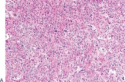
QUESTION 5.23
A. Dermatofibrosarcoma protuberans
B. Fibromatosis
C. Fibrosarcoma
D. Poorly differentiated rhabdomyosarcoma
E. Undifferentiated pleomorphic sarcoma
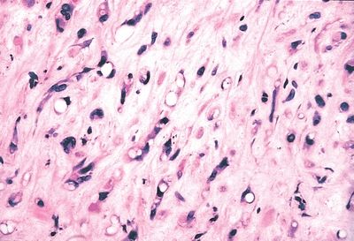
24. This photomicrograph shows the typical cellular composition of the inflammatory variant of UPS. A diagnosis of this variant can be made only if the inflammatory infiltrate consists predominantly of:
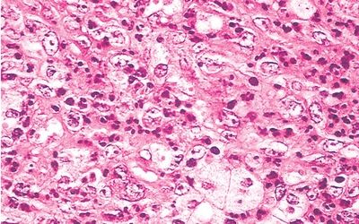
QUESTION 5.24
A. Eosinophils
B. Histiocytes
C. Lymphocytes
D. Neutrophils
E. Plasma cells
25. Which of the following variants of lipoma is frequently associated with local pain?
A. Angiolipoma
B. Angiomyolipoma
C. Chondroid lipoma
D. Fibrolipoma
E. Myxolipoma
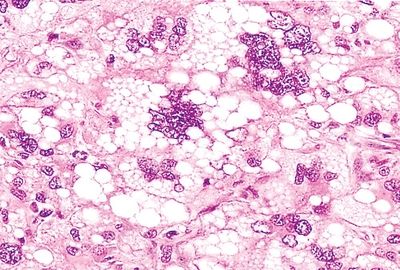
26. A 48-year-old woman undergoes excision of a gluteal fatty tumor whose typical histology is shown here. What type of adipocytic neoplasm is this?
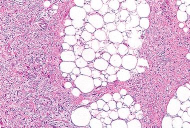
QUESTION 5.26
A. Atypical lipomatous tumor
B. Myxoid liposarcoma
C. Pleomorphic lipoma
D. Pleomorphic liposarcoma
E. Round cell liposarcoma
27. This photomicrograph shows a histologic type of liposarcoma. Which of the following is it?
QUESTION 5.27
A. Dedifferentiated
B. Myxoid
C. Pleomorphic
D. Round cell type
E. Well differentiated (atypical lipomatous tumor)
28. The vascular pattern is an important morphologic feature in the differential diagnosis of myxomatous soft tissue tumors. For example, a vascular pattern composed of dilated thick round blood vessels is typically found in:
A. Deep (aggressive) angiomyxoma
B. Intramuscular myxoma
C. Myxofibrosarcoma
D. Myxoid liposarcoma
E. All of the above
A. Dedifferentiated
B. Myxoid
C. Pleomorphic
D. Round cell type
E. Well differentiated (atypical lipomatous tumor)
30. Which of the following is a correct statement regarding leiomyosarcomas of soft tissue?
A. Histologic criteria for malignancy depend on site.
B. Most cases are associated with Epstein-Barr virus.
C. Most cases arising from large veins affect males.
D. Most cases arising in the skin affect females.
E. Superficial tumors have a worse prognosis than deep tumors.
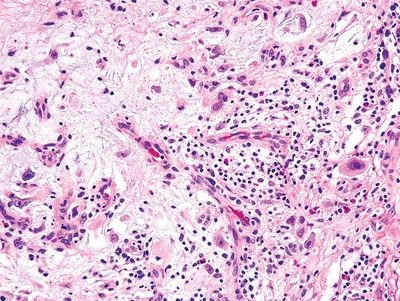
31. A highly anaplastic soft tissue neoplasm from a 5-year-old child consists of undifferentiated small cells with no distinctive features. Neoplastic cells tend to gather in dense aggregates beneath epithelial surfaces and around blood vessels, as seen in this picture. Which of the following is the most likely diagnosis?
QUESTION 5.31
A. Extraskeletal Ewing sarcoma
B. Intra-abdominal desmoplastic small cell tumor
C. Lymphoma
D. Neuroblastoma
E. Rhabdomyosarcoma
A. Alveolar
B. Botryoid
C. Embryonal
D. Pleomorphic
A. Alveolar
B. Botryoid
C. Embryonal
D. Pleomorphic
34. Which of the following forms of rhabdomyosarcoma has the worst prognosis?
A. Alveolar.
B. Embryonal.
C. Rhabdoid.
D. Pleomorphic.
E. All have equally poor prognosis.
35. The histologic appearance shown in this picture is characteristic of which type of hemangioma?
QUESTION 5.35
A. Cellular (juvenile)
B. Epithelioid
C. Hobnail
D. Microvenular
E. Sinusoid
F. Spindle cell
G. Tufted
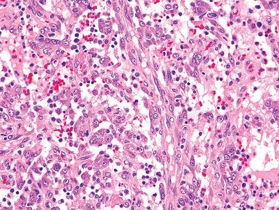
36. A 30-year-old man presents with five painful 1- to 2-cm nodules in the right lower limb. The nodules are located in the skin, subcutaneous tissue, and skeletal muscle. One of the nodules is excised and microscopically appears as the lesion shown in this picture. The tumor is immunoreactive for AE1-AE3, ERG, and CD31 and negative for EMA and CD34. Nuclear expression of INI1 is retained. Myogenic markers are negative. This clinical and pathologic presentation is diagnostic of:
QUESTION 5.36
A. Epithelioid hemangioendothelioma
B. Epithelioid sarcoma
C. Kaposiform hemangioendothelioma
D. Pseudomyogenic hemangioendothelioma
E. Retiform hemangioendothelioma
37. A 35-year-old woman undergoes resection of a large multinodular, infiltrative mass from the right lung. Its histologic appearance is shown in this picture. The tumor is diffusely immunoreactive for CD31 and ERG and focally for cytokeratin. Which of the following is the most likely diagnosis?
QUESTION 5.37
A. Angiosarcoma
B. Epithelioid hemangioendothelioma
C. Extraskeletal chondrosarcoma
D. Kaposi sarcoma
E. Metastatic signet-ring cell carcinoma
38. Which of the following vascular neoplasms develops in the upper limb as a result of chronic lymphedema secondary to radical mastectomy?
A. Angiosarcoma
B. Epithelioid hemangioendothelioma
C. Kaposi sarcoma
D. Lymphangiomyomatosis
E. Myopericytoma
39. Which of the following tumors is usually associated with tuberous sclerosis and displays HMB45 immunoreactivity?
A. Epithelioid hemangioendothelioma
B. Glomus tumor
C. Hemangiopericytoma
D. Lymphangiomyoma
E. Rhabdomyoma
Stay updated, free articles. Join our Telegram channel

Full access? Get Clinical Tree


