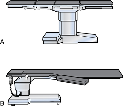
Diagnostics, specimens, and oncologic considerations

Chapter 22


Diagnostics, specimens, and oncologic considerations


Key terms and definitions


Anaplasia 


Change in cellular differentiation and orientation that causes a more primitive structural appearance and function. Anaplastic cell changes are characteristic of malignancy.


Biopsy 


Procedure for obtaining a representative tissue sample for gross and/or microscopic examination. The specimen can be obtained surgically or by other means.


Brachytherapy 


Placement of radioactive material inside of or close to a tumor. Radioactive elements are introduced through catheters inserted into the tumor.


Cancer 


Broad term that describes any malignant tumor within a large class of diseases. More than 100 different forms of cancer are known, each with histologic variations. Cancerous tumors are divided into two broad groups:


Carcinoma 


Malignant tumor of epithelial origin that affects glandular organs, viscera, and skin.


Sarcoma 


Malignant tumor of mesenchymal origin that affects bones, muscles, and soft tissue.


Chemotherapy 


Use of chemical or pharmacologic agents to treat diseases, such as cancer.


Contrast medium 


Use of a substance in the creation of density on a radiographic imaging device; radiopaque contrast injected or instilled to outline an organ or a structure. A radiolucent substance, such as air, can help define a hollow space, such as a ventricle of the brain.


Cytoreductive surgery 


Mechanical reduction in cell volume at the tumor site by sharp or blunt tissue dissection. Vessel- and nerve-sparing procedures include the use of an ultrasonic aspirator and hydrostatic pulsed lavage.


Immunotherapy 


Use of agents that stimulate or activate the body’s own host defense immune system to combat disease.


Interventional radiology 


Invasive procedures performed under radiologic control. Examples include balloon angioplasty, coronary artery stent placement, and inferior vena cava filter placement.


Neoplasm 


Atypical new growth of abnormal cells or tissues, which may be malignant or benign.


Nuclear medicine study 


Diagnostic test performed using radioactive substances to image a body part or system.


-oma 


Suffix denoting a tumor or neoplasm.


Palliation 


Measures taken to decrease the negative effects of a terminal or moribund condition. This is not considered a cure, but a temporary solution to a problematic situation. Examples include removing an obstruction or attempts to preserve fertility in the face of cancer.


Pathologic examination 


A series of tests and examinations conducted by a pathologist to determine the cause of changes in the structure or function of a body part or tissue.


Percutaneous 


Directed through the skin and tissues of the external body surface. Diagnostics, treatments, or tissue removal can be performed by direct percutaneous routes.


Plethysmography 


Procedure to determine variations in blood flow between parts.


Scintigraphy 


Recording of the emissions of radiologic substances as they are collected or secreted by tissues and/or organs in the body.


Smear 


A sample of tissue cells or fluid aspirated or scraped from a mucous membrane or a potentially pathologic site. The material is stained and studied for cellular components to make a diagnosis.


Stereotaxis 


Computerized localization of a lesion.


Tomography 


Computerized method of imaging a structure in layers.


Tumor 


Any neoplasm in which cells are permanently altered but have the capability of growth and reproduction. A tumor consists of two elements: the tumor cells themselves and a supporting framework of connective tissue and vascular supply.


Benign tumor 


Aggregation of cells that closely resemble those of the parent tissue of origin. The tumor usually grows slowly by expansion, is localized, and is surrounded by a capsule of fibrous tissue.


Malignant tumor 


Progressively growing tumor that originates from a specialized organ such as the lung, breast, or brain, or a tumor localized to a specific body system such as bone, skin, lymph nodes, or blood vessels.


Diagnosing pathology


Diagnosis of a pathologic disease, anomaly, or traumatic injury is established before a surgical procedure is undertaken. Many modalities and techniques help surgeons assess each individual patient problem, guide them through the surgical procedure, and help them verify the results of surgical intervention. The term diagnosis refers to the art or the act of determining the nature of a patient’s disease. Diagnostic procedures can be classified as follows:


Perioperative nurses and surgical technologists should be familiar with the modalities and equipment necessary to assist with diagnostic procedures in the operating room (OR) and interventional departments.


Patient care considerations for diagnostic procedures


Specimens and pathologic examination
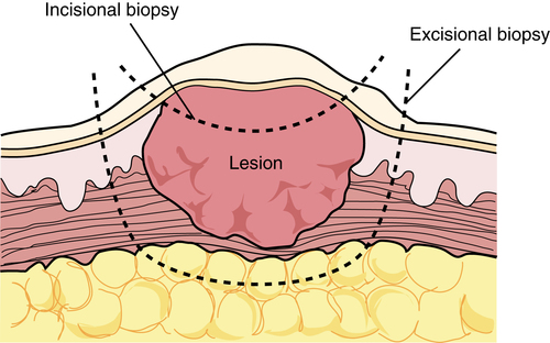

Clinical pathology is the use of laboratory methods to establish a clinical diagnosis of a disease by examining body fluid, tissue, and organs. Surgical pathology is the study of alterations in body tissues removed by surgical intervention.


Cultures and smears


Cultures


Aerobic or anaerobic cultures may be obtained before or during the surgical procedure. If obtained before the procedure the culture must be done before the skin prep. Cultures should be refrigerated or sent to the laboratory immediately. The culture tube and plastic transport bag should be labeled with the patient’s identification, date, and site of culture.


The sterile culture tube and swab are prepackaged in a peel pouch and are dispensed to the sterile field. The culture tube has a small sealed plastic media chamber at the bottom that preserves the material obtained from the patient on the swab. Nothing other than the fluid or tissue to be cultured should touch the sterile swab.


The sterile swab is withdrawn from the tube by the sterile team member and dipped into the area to be cultured. The swab is replaced into the culture tube and seated tightly. The media chamber at the bottom of the tube is crushed to release the culture media and immerse the tip of the swab. The scrub person can drop the closed culture tube into a small plastic bag held by the gloved circulating nurse, who will affix the patient’s identification label and biohazard sticker.


Cultures for suspected anaerobic microorganisms require immediate attention. Exposure to room air may kill anaerobes in a few minutes. The purulent material can be aspirated into a sterile disposable syringe through a disposable needle. This needle is removed with a hemostat and placed with counted sharps on the instrument table.


Air is expelled away from the field, and the syringe is capped with the syringe tip supplied with the syringe or a Luer Loc cap; the syringe is then sent immediately to the laboratory in a plastic bag with a biohazard sticker affixed to the outside. The syringe should not be sent to the laboratory with the needle attached, and the needle should not be recapped by hand because of the potential for a needlestick injury.


Biopsies


The removal of tissue or fluid for diagnosis is referred to as a biopsy. Biopsy specimens can be obtained by many invasive methods. The pathologist determines and/or confirms the diagnosis by histologic examination (the study of tissue) and cytologic analysis (the study of cells).


Frozen section


Special preparation and examination of tissue can determine whether it is malignant and whether regional nodes are involved. When the surgeon removes a piece of tissue and wants an immediate diagnosis, it is placed in a basin or specimen container without any added preservative, such as formalin or normal saline solution. Formalin or normal saline solution will alter the freezing process used in the specialized pathologic study. The circulating nurse should alert the pathologist that a frozen section will be sent. When the tissue examination is complete, the pathologist will report the results directly to the surgeon in the OR.


The patient’s level of consciousness should be considered during report of pathologic results, especially if the patient is awake. The use of speakerphones should be avoided for patient confidentiality. If a malignant lesion is present and the individual situation calls for it, the surgeon proceeds with a radical resection of the affected organ or body area. In some situations, additional tissue is needed for diagnosis.


Permanent section


The specimen is placed in fixative, commonly formalin, for several hours to cause the cells to become firm. The fixed specimen is placed in a machine that removes all of the water from the tissues, replacing it with paraffin. When this process is complete, the specimen is embedded into a block of wax. It is placed in a microtome and is sliced tissue-paper thin and floated in a bath of water. The slices are placed on glass slides, where the paraffin is dissolved with solvent and the water is restored to the tissue on the slide. The slices readily accept dyes and stains used for diagnosis. Permanent section is the best diagnostic biopsy tool. Box 22-1 lists several tissue dyes and stains used in diagnostic procedures.


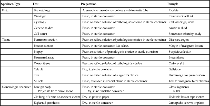
Collecting surgical specimens


All tissue removed during the surgical procedure is sent to the pathology laboratory for verification of the diagnosis. Any unidentifiable or unusual item removed from a patient should be sent for identification by the pathology department. Some examples include removal of retained surgical items, such as instruments, sponges, and towels. Any questionable item should be sent to the lab and documented by the circulating nurse. Facility policy and procedure should delineate disposition of all surgical specimens.


Tissue specimens may be stored in a refrigerator in the laboratory or some other location within the OR suite until they are taken to the pathology department at the end of each day’s schedule of surgical procedures or at intervals during the day.


Correct solutions for storage and correct labeling of specimens are critical for accurate diagnosis. Table 22-1 lists types of specimens for pathologic study.


Specimen containers may be plastic containers, waxed cardboard cartons, or glass jars with preservative solution. AORN recommended practices suggest that the specimen container be part of the sterile setup so the container can be closed on the field, minimizing the handling of biohazardous material. The closed, labeled container is placed into a plastic bag or additional container with a biohazard sticker for transport to the laboratory. The double-packaged specimen should only be handled while wearing gloves.


The handling of specimens should be kept to a minimum and only while wearing gloves and appropriate personal protective equipment (PPE). Use care to avoid contaminating the outside of a specimen container. If it is contaminated, wipe it with an antiseptic solution. Always wash hands thoroughly after removing gloves that have been worn to handle specimens. If instruments are used for handling, be careful not to tear, crush, or damage tissue. The routine care for each type of tissue specimen may vary as follows:


• Pathology tissue specimens should not be allowed to dry out. Saline or a solution of aqueous formaldehyde (10% formalin) is commonly used as the fixative until the specimen is processed further in the laboratory. Formalin is flammable and should not be stored in the OR. The formalin is added after the specimen is in the container. Dropping the specimen into a prefilled container can cause a splash.


• Stones are placed in a dry container so they will not dissolve. Organs containing stones, such as the gallbladder, may be placed in saline or formalin.


• Foreign bodies should be sent for accession according to policy, and a record is kept for legal purposes. The description and the disposition of the object are recorded. A foreign body may be given to the police, surgeon, or patient, depending on legal implications, policy, or the surgeon’s wishes. A chain of specimen custody slip should be signed by all persons handling the specimen.


• Forensic evidence, such as bullets or knife blades, should be placed in a dry plastic container. Do not allow the item to contact metallic basins because ballistic evidence could be altered. Chain of custody documentation should accompany the specimen at all times to protect the evidence.


• Amputated extremities are wrapped in plastic before sending them to a refrigerator in the laboratory or morgue. Avoid placing the amputated limb in the patient’s field of vision. Most patients needing amputation have spinal or epidural anesthesia for the procedure. The sight of the body part may cause emotional distress in the patient. The patient may request that an amputated extremity be sent to a mortuary for preservation for burial with his or her body after death. This request must be noted on the requisition sent to the laboratory. Refer to institutional policy and procedure for the care of amputated limbs.


Radiologic examination


An x-ray is a high-energy electromagnetic wave capable of penetrating various thicknesses of solid substances and affecting photographic plates. X-rays are generated on a vacuum tube when high-velocity electrons from a heated filament strike a metal target (anode), causing it to emit x-rays.


The image obtained by the use of x-rays may be referred to as an x-ray, roentgenogram, radiograph, or other -gram or -graph name associated either with the specific technique used to obtain the photograph or the anatomic structures identified (e.g., mammogram, x-ray, or radiograph). Most facilities have eliminated the use of x-ray film and have changed to a digital format.


X-rays are also used for diagnostic imaging with fluoroscopy, computed tomography (CT), and digital radiography. Computerized radiologic images may be stored on diskettes.


Types of radiologic equipment and accessories


Many hospitals have one or more rooms within the OR suite that are equipped for diagnostic as well as intraoperative radiologic procedures. In some facilities, OR personnel are cross-trained to assist with diagnostic and interventional procedures in the radiology department.


Contrast media


Agents composed of nonmetallic compounds or heavy metallic salts that do not permit passage of radiant energy are radiopaque. When exposed to x-rays, the lumina of body structures filled with these agents appear as dense areas. Radiopaque contrast media frequently used for the procedures to be described are listed in Box 22-2.


Studies have shown that contrast agents are excreted in breast milk in small amounts. Contrast agents cross the placenta and are excreted by the fetal kidneys into the amniotic fluid. Sensitization of the infant has not been shown. The contrast is cleared by the mother’s circulation within 24 hours. She may wish to abstain from breast feeding during that period. Refer to the product package insert for specific information.


Some contrast agents are fluorescent dyes. Most contain iodine, either bound or unbound. The contrast media is designated low-osmolarity or high-osmolarity. Low-osmolarity is associated with fewer reactions to contrast.12,6,9,10 Few patients have an immediate reaction; however, a small population of patients may react several hours later. Known reactors may be given prednisone and Benadryl several hours before the anticipated use of any contrast agents.9,10


A history of systemic sensitivity to substances that contain iodine, such as shellfish, or other allergies is obtained before these agents are injected.2,8 True shellfish allergies are not related to iodine, but to specific proteins in the fish. True allergy is not a common event.2,5,9 Studies have shown that other allergies, such as milk and egg, both high in protein, may play a role. Precautionary administration of prednisone and Benadryl minimizes the risk. The use of low-osmolarity contrast helps to decrease reactions.9


Iodine allergy information may be unreliable, because studies have shown that immediate reactions to contrast are nonimmunologic in nature.5,9 This means that the adverse reaction is not caused by allergy. The patient’s Ig-E immunoglobulin is not activated. True allergic reactions to any contrast agent ingredient stimulate Ig-E immunoglobulin and the patient can exhibit hives, angioedema, respiratory difficulty, and cardiovascular collapse.1,2,9


A test dose of 1 or 2 mL may be given before the dose required for x-ray study to rule out a potential reaction. The patient should be observed for symptoms of reaction throughout the procedure. Signs of reaction may include the following:


Radiologic table


The tabletop the patient lies on is made of acrylic or some other radiolucent material (Fig. 22-2). Some operating beds have a Bakelite top that fits over the length of the table and permits insertion of the x-ray cassette at any area. For fluoroscopy with image intensification, the entire top must be radiolucent because the image intensifier is positioned underneath the table. If the entire operating bed is not radiolucent, the foot section can be lowered and a radiolucent extension attached that will accommodate specialized x-ray machinery.


Portable x-ray machine


An x-ray tube mounted on a portable electric- or battery-powered generator of a nonexplosive design may be moved from one room to another in the OR suite and postanesthesia care unit (PACU). A portable x-ray machine offers the advantages of flexibility in scheduling procedures and availability for when and where it is needed.


Portable x-ray machines can be a source for cross-contamination. All portable equipment should be thoroughly disinfected before being brought into the OR/PACU and again after use. It should be stored within the perioperative environment between uses. Portable x-ray machines are used for intraoperative angiography, cholangiography, orthopedic localization, and urologic contrast procedures.


Intraoperative images require the x-ray machine to be in proximity to the sterile field and could cause contamination. A few options to prevent contamination include draping the machine with a specialized drape or temporarily placing a sterile towel over the site to be imaged. Extreme care is taken to prevent contamination when the towel is removed. Reasons for intraoperative x-ray imaging include performing diagnostic examinations with contrast media or looking for a lost surgical object.


In the PACU the portable x-ray machine is used for immediate (stat) chest and KUB/flat plate films. X-ray films can be used to check the location of implants and delayed passage of contrast medium through an organ system.


Fluoroscope


Similar to an x-ray generator, a fluoroscope has an additional screen, composed of fluorescent crystals, which lies in contact with a photocathode. When an x-ray beam passes through this screen, it fluoresces. Fluorescent light sets electrons free from an adjacent photocathode to produce an electron image. Rather than this image of body structures being photographed on x-ray film, it is reproduced as a digital image on a luminescent screen. Known as fluoroscopy, examination under a fluoroscope allows visualization of both form and movement of internal body structures. Fluoroscopy is used frequently for both preoperative and intraoperative procedures.


It is an invasive technique, because a fluorescent substance must be injected or a radiopaque device inserted. When exposure time is prolonged to perform a procedure with visual fluoroscopic control (e.g., cardiac catheterization), fluoroscopy exposes the patient and personnel to radiation at higher levels than do conventional x-rays. Personnel must wear lead aprons during fluoroscopy, even though a lead shield is part of the installation. Patients in the vicinity should be protected with gonadal and thyroid shields.


C-arm image intensifier


Designed primarily for orthopedic procedures, foreign body and calculi localization, and catheter placement, mobile image intensifiers offer the same advantages and disadvantages as do portable x-ray machines. The C-arm, so named because of its shape (see Fig. 13-2), keeps the image intensifier and x-ray tube in alignment; the intensifier is directly under the tube. It can be moved from an anterior to a lateral position. Utility of the mobile C-arm image intensifier is enhanced when the system is capable of making electronic x-rays for permanent records. An additional formatting device is required for this function.


Computerized digital subtraction processor


After IV injection of a radiopaque contrast medium, the computerized digital subtraction x-ray imaging system visually records perfusion within the cardiovascular system (e.g., extracranial and intracranial vessels).


Initially, before contrast medium is injected, fluoroscopic body images are converted to digital data for storage in a memory unit in the processor. Termed the mask image, these digitized data are integrated into single or multiple video frames. The video signal is logarithmically amplified and digitized. The mask image is electronically subtracted from subsequent images with the contrast medium. This process removes unwanted background, thus providing optimal visualization of vessels with contrast density that cannot be achieved by other image intensifiers. The resultant images (digital x-rays) are displayed on a video screen and can be recorded on diskette or videotape.


Radiologic diagnostic procedures


Chest x-ray


Most surgeons consider a chest x-ray film as an extension of the patient’s history and physical examination if it is clinically necessary. An x-ray study of the chest may be part of the admission procedure for elective surgical patients to rule out unsuspected pulmonary disease that could be communicable or would contraindicate the use of inhalation anesthetics.


This procedure may be routine for patients older than 40 years. It is always a part of the diagnostic workup in patients with suspected or symptomatic pulmonary abnormalities if they will be undergoing general anesthesia.


Mammography


A technique for projecting an x-ray image of soft tissue of the breast, mammography is the most effective screening method for early diagnosis of small, nonpalpable breast tumors. Mammography may be performed in conjunction with ultrasonography if the woman has fibrocystic breasts and difficult-to-palpate masses less than ¼ inch (0.5 cm) in diameter.


Three views of each breast are exposed to conventional x-rays. The procedure may be somewhat painful for the woman, because compression is needed for radiologic imaging. Tumors appear on the mammogram as opaque spiculated (star-shaped) areas or, occasionally, as areas of calcification. The radiologist places a small, BB-like radiolucent bead with crosshairs over a suspicious area. Another x-ray film of this area is obtained. Fine needle aspiration may be performed under ultrasonic guidance to gather cells for cytologic study.


Patient teaching.

Women 35 to 40 years of age should have a baseline screening mammogram. Between the ages of 40 and 50 years, they should have additional screening films yearly or every other year as recommended by their physician. After the age of 50 years, every woman should have a yearly mammogram.


Mammography is not contraindicated in women with breast implants. The technician obtaining the x-ray should be informed of previous breast surgeries, hormone replacement therapy, and augmentation mammoplasty. Special breast-imaging techniques are employed to displace an implant or obtain an x-ray film of extremely dense breast tissue.


Men with suspicious breast lesions or pronounced gynecomastia should have mammographic screening to rule out cancerous tissue.


Every patient should be taught breast self-examination and practice it every month as a baseline diagnostic test. Early diagnosis is the best chance for a cure. Figure 22-3 depicts the average sizes of masses discovered in patients who do and do not practice breast self-examination.


Computed tomography


In computed tomography, special complex and expensive equipment uses an x-ray in conjunction with a computer. Because the x-ray beam moves back and forth across the body to project cross-sectional images, the technique is referred to as computed tomography (CT), computed axial tomography (CAT), or simply scanning. It produces a highly contrasted, detailed study of normal and pathologic anatomy.


The x-ray tube and photomultiplier detectors rotate slowly around the patient’s head, chest, or body for 180 degrees in a linear fashion along the vertical axis. The computer processes the data and constructs a three- or two-dimensional picture on a black-and-white monitor or in colors that correspond to the density of tissue. Structures are identified by differences in density. This picture is photographed for a permanent record. The computer also prints out on a magnetic disk the numerical density values related to the radiation-absorption coefficients of substances in the area scanned. The radiologist uses this printout to determine whether a substance is fluid, blood, normal tissue, bone, air, or a pathologic lesion. The exact size and location of lesions in the brain, mediastinum, and abdominal organs are identified.


CT becomes an invasive procedure when a radiopaque contrast medium is injected IV to enhance visualization of the vascular and renal systems. Oral diluted barium contrast can be used to examine the gastrointestinal tract when a perforated organ is not suspected. Tissue biopsy can be performed under CT guidance. Needle aspiration can be performed under direct CT visualization.


CT exposes the patient to ionizing radiation and a potential allergic reaction to contrast medium, if used. To ensure proper use of this complex equipment, as well as to protect the patient from excessive or unnecessary radiation, the procedure is done under the supervision of a qualified radiologist.


Ventriculography


Ventriculography is the x-ray study of the ventricles after injection of gas directly into the lateral ventricles of the brain. It may be used to evaluate a patient with signs of increased intracranial pressure as a result of blockage of cerebrospinal fluid circulation. Ventricular needles or catheters are inserted into one or both lateral ventricles through holes made in the skull. The ventricular needle has a blunt, tapered point that prevents injury to the brain as it is inserted into the ventricle. Openings on the side near the point permit removal of spinal fluid and injection of gas. If the patient is an infant whose suture lines in the skull are not yet closed, needles are inserted through these.


The entire ventriculographic procedure may be done in the OR. Otherwise, the holes are drilled and the ventricular needles or an intraventricular catheter is inserted and the patient is then transferred to the radiology department or an interventional room within the OR suite. If a lesion is identified, the diagnostic procedure may be followed immediately by a surgical procedure because of the possibility of a further increase in intracranial pressure. If the patient is returned to the unit after the procedure, sterile ventricular needles should accompany the patient. If intracranial pressure becomes too great after gas injection, a needle can be inserted to remove the gas.


Angiography and arteriography


Angiography is a comprehensive term for studies of the circulatory system after injection of a radiopaque substance to permit visualization of the venous blood vessel system. These procedures are useful in the differential diagnosis of arteriovenous malformations, aneurysms, tumors, vascular accidents, or other circulatory abnormalities caused either by traumatic injury or an acquired structural disease.


Intraoperative studies often are essential to assess the results of vascular reconstruction. Angiography is one method of assessment to confirm the position and patency of an arterial or venous graft or the quality of a restored vessel lumen. Intraoperative angiography frequently is indicated for these assessments in the peripheral vessels of extremities. After insertion of a bypass graft or endarterectomy, patency of the graft or vessel is checked by pulsations and also by arteriography to examine the arterial blood vessels of the circulatory system.


Angiography is also used at the time of the surgical procedure to identify vascularity or the exact location of some types of lesions in the extremities, brain, and thoracic and abdominal cavities. After venous injection of radiopaque contrast medium, radiologic studies are done. Techniques and equipment to be used will vary according to the specific procedure, but all types of angiography have the following features in common:


1. Access to the vessel (either an artery or vein) to be injected with a radiopaque contrast medium may be made by a percutaneous puncture or a cutdown. An IV drip is maintained when the latter approach is used.


a. Cannulated needles with or without a radiopaque plastic catheter, similar to those used for IV infusions, may be used for a percutaneous puncture to penetrate an artery or vein. To prevent backflow of blood, cannulated needles have an obturator that remains in place until the contrast medium is injected. Long catheters have a guidewire to assist threading through the vessel.


b. A Seldinger (18-gauge) needle has a sharply beveled inner cannula and a blunt outer cannula. The blunt end of the outer cannula prevents trauma to the vessel. After insertion, the inner cannula is replaced with a guidewire and the outer cannula is removed. A 20-cm vessel dilator sheath (which is approximately 8 inches long) is threaded over the guidewire to create a track for a radiopaque catheter.


Stay updated, free articles. Join our Telegram channel

Apr 6, 2017 | Posted by in GENERAL SURGERY | Comments Off on Diagnostics, specimens, and oncologic considerations

Full access? Get Clinical Tree

Get Clinical Tree app for offline access