Developmental, Congenital, and Childhood Diseases and Disorders


Developmental, Congenital, and Childhood Diseases and Disorders


Learning Objectives


After studying Chapter 2, you should be able to:


1. List the possible causes of congenital anomalies.


2. Discuss the purpose and procedure of amniocentesis.


3. Discuss genetic disorders and syndromes.


4. Trace fetal circulation.


5. Describe the condition of prematurity and associated disorders: the causes and treatment.


6. List possible risk factors of retinopathy of prematurity.


7. List symptoms and signs of Down syndrome.


8. List and discuss diseases of the pediatric nervous system.


9. Distinguish between muscular dystrophy and cerebral palsy.


10. Describe patent ductus arteriosus.


11. Name and describe the most common congenital cyanotic cardiac defect.


12. List and discuss musculoskeletal conditions of newborns and young children.


13. List and discuss pediatric genitourinary conditions.


14. List and discuss pediatric diseases of the digestive system.


15. List the major clinical manifestations of cystic fibrosis.


16. Distinguish between Klinefelter’s syndrome and Turner’s syndrome.


17. Describe the clinical condition of congenital rubella syndrome.


18. List and discuss contagious diseases of children.


19. Discuss possible causes and prevention of SIDS.


20. Distinguish between croup and epiglottitis.


21. Describe the symptoms, signs, and treatment for tonsillitis.


22. Discuss the treatment of asthma.


23. List types of worms that may infest the GI tract.


24. List the symptoms and signs of anemia; describe the pathology of leukemia.


25. Explain the etiology of erythroblastosis fetalis.


26. Name some warning signs of lead poisoning.


27. Describe the infant born with fetal alcohol syndrome.


28. Discuss causes and treatment of bronchiolitis.


Developmental and Congenital Disorders


Developmental Characteristics and Congenital Anomalies


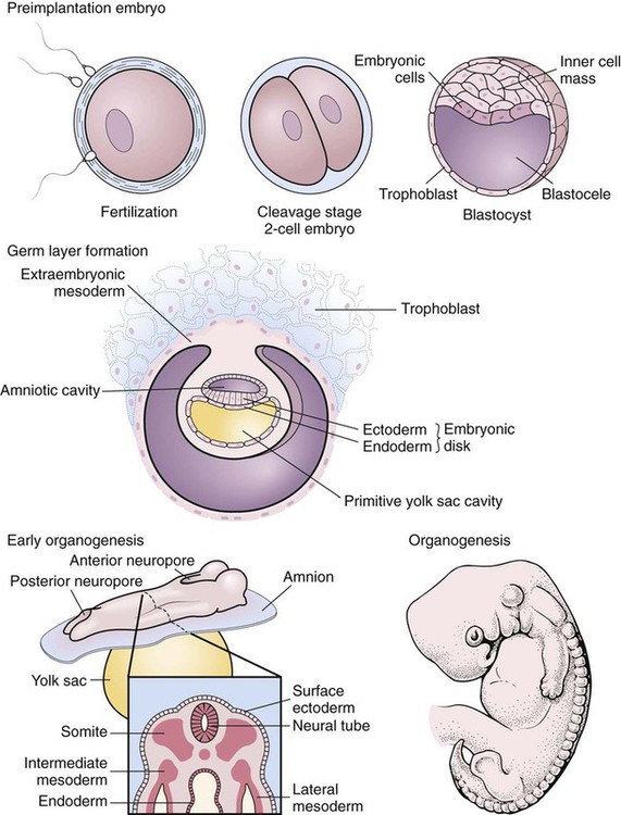
The developmental process begins with conception and progresses as a gradual modification of the structure and characteristics of the individual (Figure 2-1). The first 2 months of the gestational period is considered the embryonic period, after which the developing human being is considered a fetus. At any point in this prenatal development, during the birth process (perinatal period), or during the neonatal and postnatal periods, the development may diverge from normal, generating a developmental dilemma. Causes of these dilemmas can be many or even unknown. Pregnant women are encouraged to abstain from smoking, consuming alcohol, and taking any form of medication or drugs without their physician’s knowledge and consent and to prevent any situation that may expose the developing fetus to toxic substances. Table 2-1 describes the specific stages of development during this important period of life. E2-1


TABLE 2–1


Monthly Changes During Prenatal Development


END OF MONTH* SIZE OF EMBRYO OR FETUS DEVELOPMENTS DURING THE MONTH
 1 6 mm Arm and leg buds form; heart forms and starts beating; body systems begin to form.
 2 23-30 mm, 1 g Head nearly as large as body; major brain regions present; ossification begins; arms and legs distinct; blood vessels form and cardiovascular system fully functional; liver enlarges.
 3 75 mm, 10-45 g Facial features present; nails develop on fingers and toes; can swallow and digest amniotic fluid; urine starts to form; fetus starts to move; heartbeat detected; external genitalia develop.
 4 140 mm, 60-200 g Facial features well formed; hair appears on head; joints begin to form.
 5 190 mm, 250-450 g Mother feels fetal movement; fetus covered with fine hair called lanugo hair; eyebrows visible; skin coated with vernix caseosa, a cheesy mixture of sebum and dead epidermal cells.
 6 220 mm, 500-800 g Skin reddish because blood in the capillaries is visible; skin wrinkled because it lacks adipose in the subcutaneous tissue.
 7 260 mm, 900-1300 g Eyes open; capable of survival but high mortality rate scrotum develops; testes begin their descent.
 8 280-300 mm, 1400-2100 g Testes descend into the scrotum; sense of taste is present.
 9 310-340 mm, 2200-2900 g Reddish skin fades to pink; nails reach tips of fingers and toes or beyond.
10 350-360 mm, 3000-3400 g Skin smooth and plump because of adipose in subcutaneous tissue; lanugo hair shed; fetus usually turns to a head-down position; full term.

*These are 4-week (28-day) months.


From Applegate EJ: The anatomy and physiology learning system, ed 3, Philadelphia, 2006, Saunders.


Congenital Anomalies


Congenital anomalies can be mental or physical and can vary widely in severity, from trivial to fatal. They are present at birth but might not be detected until later in infancy or childhood. The limbs or organs may be malformed, duplicated, or entirely absent. Organs sometimes fail to move to their proper location or fail to open or close at the right time. Anomalies are seldom isolated and are likely to occur in multiple forms and/or organs or organ tissues.


The cause of congenital defects may be genetic, nongenetic, or a combination of both. Nongenetic causes include infection in the mother, drugs taken by the mother, the age of the mother, radiographic examination made early in pregnancy, or injury to the pregnant woman or the fetus. The cause often is unknown. However, prenatal care and advanced surgical techniques have greatly improved the management of anomalies that are compatible with life. Parents of a special-needs child face emotional and physical challenges and deserve medical attention. A team approach is ideal, including medical assessment by physicians and appropriate therapy for the child, and family involvement and participation in a support group for the parents.


Genetic Disorders and Syndromes


Genetic syndromes are a form of congenital anomalies. Genetic information is contained in microscopic threadlike structures in the nucleus of human body cells. The genetic material contained within the genes is responsible for inheritance traits. Each cell within the human body contains 46 chromosomes arranged in 23 pairs. Twenty-two pairs of the chromosomes are termed homologous pairs and the remaining pair is the sex chromosomes.


Genetic disorders and syndromes are the result of an abnormal gene taking up residence on one of the 22 pairs of nonsex chromosomes. The 22 pairs of homologous pairs are also identified as autosomes. When the abnormal gene that causes the condition is on one of the 22 pairs of autosomes, the condition is termed an autosomal inherited condition.


Some of the autosomal inherited conditions have a dominant pattern of inheritance; other conditions may be the result of autosomal recessive traits. Dominant inheritance occurs when only one gene of the pair has the ability to produce symptoms, thus making it dominant over the normal gene. The parent who carries the abnormal gene has a 50% chance of transmitting the defective gene to each of his/her offspring. When the condition is considered as autosomal recessive inheritance, the parent’s abnormal genetic makeup must have genetic errors on both of the genes of the pair resulting in the carrier usually having no symptoms of the disorder. The autosomal recessive disorder has a 25% chance of surfacing when two carrier parents conceive a pregnancy. The child could be affected by the autosomal inherited condition when the pair of affected genes are present.


Methods of Prenatal Diagnosis


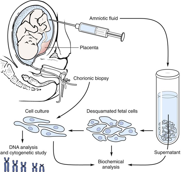
One can diagnose congenital anomalies in a fetus by taking a fluid sample from the amniotic sac between the 15th and 18th weeks of pregnancy. This procedure, known as amniocentesis, allows amniotic fluid to be tested and cells to be microscopically examined for abnormal substances or chromosomal abnormalities. An example of an abnormal substance is an elevated alpha-fetoprotein (AFP) level. Amniocentesis is not without risk to the mother and baby.


Abnormalities of the spine, skull, and many organs such as the heart and kidneys may be discovered during ultrasound studies of the fetus. An alternative procedure called chorionic villus biopsy (CVB) can be performed in the second month of pregnancy. The gynecologist, guided by ultrasound, directs an instrument toward the placenta in the womb and obtains a tissue sample. The safety of this procedure has not been proven, and some data link this test to limb abnormalities.


Many, but not all, congenital disorders can be detected through prenatal diagnosis (Figure 2-2).


The diagnosis of potential genetic or neural tube deficit disorders may provide the future parents with time to obtain counseling and to prepare for the many health needs of the infant before the child’s birth. When the testing indicates a potential abnormality, the delivery and nursery staff is provided with the opportunity to prepare for the delivery. E2-2


image Enrichment


Conjoined Twins


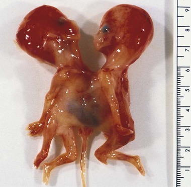
During the conception process, the fertilized egg—the embryo—may divide, creating identical twins. Conjoined twins result when the separation process of identical twins fails to complete before the 13th day after fertilization


As with identical twins, the embryo originates from a single fertilized ovum and occupies one placenta. For an unidentifiable reason, however, the normal separation of the embryo into twins stops before completion, resulting in a partially separated embryo that continues to mature into conjoined fetuses. Conjoined twins occur more often in female embryos than in male embryos and result in two fetuses that are joined at some point on their bodies. More of these children are being born alive as a result of specific prenatal diagnosis and surgical intervention to facilitate the delivery.


These children may be joined at different locations of the body and may share various organs. The attachment to each other may involve a small portion of tissue or may be as extensive as fusion at the head or sharing of an organ or body part. Common types or variations usually are categorized by the location and involvement of the junction through the term pagus, meaning fastened, included in the classification terminology.


Twins with a cranial union are considered to be craniopagus. Those with anterior junction at the chest, often sharing the heart and vital portions of the chest wall and internal organs, are called thoracopagus conjoined twins. The term pygopagus describes those twins who are joined posteriorly at the rump. Another posterior junction occurring at the sacrum and coccyx is termed ischiopagus. When the connection proceeds from the breastbone to the waist, the term omphalopagus describes the junction. A very rare form, dicephalus, is the condition in which the individual has one body and two separate heads and necks.


Modern technology and medical advances have recently helped physicians and surgical teams to successfully separate some of these twins. In some separation procedures, one or both of the children have died during or shortly after the surgery. The children and their families require emotional support and education about the possible outcomes of the condition. E2-3


Prematurity


Preterm Birth or Prematurity


Description

Preterm birth or prematurity is the result of birth before the 37th gestational week of pregnancy. The condition of prematurity describes the birth of a low-weight, underdeveloped, and short-gestation infant and is considered the leading cause of death during the neonatal period. These high-risk infants are born with incomplete development of organ systems. These infants are at a greater risk than full-term infants for developing serious health problems including: cerebral palsy, mental retardation, chronic lung disease, gastrointestinal problems, vision and hearing loss, and multisystem developmental delays. E2-4


image ICD-9-CM Code 765.00 (Extreme immaturity)


image ICD-10-CM Code P07.00 (Extremely low birth weight newborn, unspecified weight)


image ICD-9-CM Code 765.10 (Other preterm infants)


image ICD-10-CM Code P07.00 (Extremely low birth weight newborn, unspecified weight)


Weeks of Gestation:


image ICD-9-CM Code


image ICD-10-CM Code


Symptoms and Signs

Premature babies may weigh down to 12 ounces or less at the time of birth. Their physical development is at various stages depending on the length of gestational time. The smaller of these infants has little subcutaneous fat, palms and soles with few creases, possible undescended testes in the male, and a prominent clitoris in the female. Many of these very tiny and immature babies lack the ability to suck or swallow or have weakened sucking or swallowing reflexes. The lungs are often underdeveloped, leading to respiratory dangers. The immature neurologic system can lead to some difficulties in maintenance of body temperature, problems in controlling cardiac function, spontaneous episodes of apnea, or may result in seizure activity. An immature immune system makes the risk of infection high.


Etiology

There are many reasons that these infants enter the world before reaching the traditionally accepted gestational age of 40 weeks and thus have very low birth weights. Causes of premature labor resulting in a premature infant are an incompetent cervix, bicornate uterus, toxic conditions, maternal infection, trauma, premature rupture of the amniotic membranes, history of previous miscarriages, multiple gestations, intrauterine fetal growth retardation, and other physical conditions of the mother, such as pregnancy-induced or chronic hypertension. Diabetes, heart disease, kidney disease, poor nutrition, substance abuse, and lack of prenatal care also contribute to the incidence of the mother giving birth to a preterm infant. In some cases, the etiology is never identified. Attempts may be made to halt premature labor by having the mother on complete bedrest or using drug therapy to slow or halt contractions. The mother is often given a short course of steroids to aid fetal lung maturation. Many times the efforts to allow the fetus more time to grow and mature in the mother’s uterus are unsuccessful.


Treatment

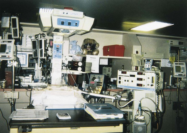
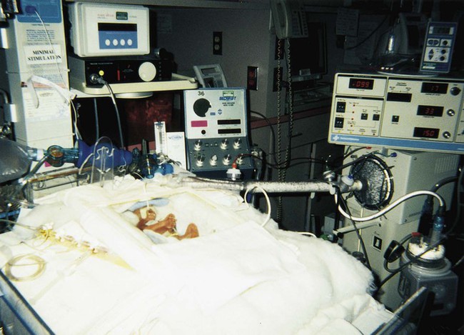
Treatment varies depending on the gestational age, weight, present or subsequent conditions, anomalies, and nutritional status. Intravenous (IV) fluids and hyperalimentation are necessary to encourage growth and development of the premature infant. Airway management and pulmonary functioning are monitored very closely. Many of the smallest babies are intubated endotracheally, and respiration is maintained by mechanical ventilation. Recent advances in respiratory care for tiny premature infants permit extubation of the infant and maintenance of the airway by continuous positive airway pressure (CPAP) through the nose. Pulse oximeters constantly monitor oxygen (O2) saturation levels and heart rate. Body temperature is monitored closely and maintained at normal levels (Figure 2-4). When infection is a risk due to maternal prenatal infection or extended time of ruptured amniotic sac, vigilance is required for the onset of any symptoms of infection and antibiotics are often started presumptively. When early signs of infection occur, aggressive treatment is introduced. Monitoring of blood glucose, blood pressure, and body temperature are performed on a frequent and regular basis and any treatment necessary is instituted to maintain optimal levels.


Comfort measures include placing an article with the mother’s scent in the isolette or crib with the infant. These tiny babies are swaddled to simulate the closeness and feeling of the womb. Lights are kept at a dim level and harsh auditory stimuli are kept at a minimum.


Prognosis

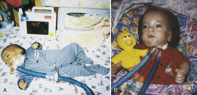
Advances in technology have made survival of low-weight and short-gestation infants possible. The prognosis for these children varies depending on gestational age, weight, and the occurrence of anomalies and developmental deficits. There are documented cases of 12-ounce and/or 22-gestational-week babies surviving. They fall into the 1% of premature babies born at that weight and gestational age. Being born before the normal prenatal development is complete, these children often have many problems to overcome (Figure 2-5).


A primary risk is a cerebral bleed, which may occur during the labor and delivery process or may result from handling after delivery. The cerebral bleed may cause the development of cerebral palsy, mental functioning deficiencies, or other neurologic conditions.


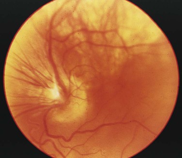
Another major concern is underdevelopment of the pulmonary system, including the lung tissue and the airway. Some pulmonary conditions these infants experience are infant respiratory distress syndrome (IRDS), bronchopulmonary dysplasia (BPD), laryngomalacia, tracheomalacia, and bronchomalacia. Lack of body fat can affect the maintenance of body temperature. Any stress or increased or high supplemental O2 flow may be responsible for retinopathy of prematurity (ROP) and possible blindness. Necrotizing enterocolitis (NEC) is a danger in the digestive system because of the reduced tolerance of the alimentary tract. Atrial septal defect (ASD) and patent ductus arteriosus (PDA) often are present because the fetal circulatory system has failed to mature.


The generally accepted gestational age for 50% of infants to survive the birth process and perinatal period is 24 weeks, with a greater percentage of infants surviving as gestational age increases. Improvements in technology are making it possible for more and more of these tiniest infants to survive (Figure 2-6). E2-5


image
FIGURE 2–6 A, Same premature infant from Figure 2-5 at age 9 months; weight 11 lb, 8 oz. B, Same infant at age 10 months, weight 13 lb. (Courtesy David L. Frazier, 1999.)


Infant Respiratory Distress Syndrome


Patient Teaching

Give the parents printed information about the disease. Parents will need emotional support and understanding. They should be referred to community support groups. Allow the parents to verbalize their anxieties and fears to a compassionate and understanding individual, thus providing a validation of their situation.


Unfortunately, the occurrence of IRDS, along with its treatment modalities, often predisposes premature infants to the development of bronchopulmonary dysplasia (BPD).


Bronchopulmonary Dysplasia


Treatment

The goal of treatment is replacement of the damaged alveoli. Children grow new alveoli until about 8 years of age. Infants who have BPD need to grow new alveoli to replace those damaged by scarring. As this replacement occurs, the severity of the condition lessens. Supportive treatment includes supplemental oxygen and adequate nutritional support. The types of medications used include diuretics and bronchodilators, including beta2-agonists, anticholinergic drugs, and theophylline. Antiinflammatory drugs such as steroids also may help.


Supplemental O2 therapy may be needed for several weeks, occasionally for more than 1 year. This therapy usually is by nasal canula; however, if the infant has a tracheostomy, it may be delivered by a tracheostomy collar or by CPAP. O2 saturation levels must be monitored with a pulse oximeter so they can be maintained at 90% or greater. The pulse oximeter also monitors heart rate. As the infant grows and matures, blood oxygen saturation levels may be maintained on room air, usually by the age of 1 year.


Diuretics help to reduce fluid accumulation in the lungs and reduce the incidence of pulmonary hypertension and right-sided heart failure. Bronchodilators are administered to reverse the narrowing of the bronchi resulting from inflammation or bronchospasm, thus allowing more oxygen to reach the lung tissue. These drugs may be administered orally as syrups or by aerosol inhalation. The antiinflammatory drugs help to prevent the inflammatory process from becoming severe.


Adequate nutrition is needed for the infant’s growth and to meet the increased caloric demand resulting from difficult breathing. High-calorie formulas are used. The infant must be held with the head raised slightly and the formula given frequently in small amounts to prevent gastroesophageal reflux disease (GERD). Some infants are given medications to prevent GERD and other medications, such as antacids or histamine-2 blockers, to reduce gastric acid.


Prognosis

Prognosis is good with early and aggressive intervention, prudent monitoring, and maintenance of adequate oxygen saturation levels and heart rate. Resolution of the condition is slow, and improvement is gradual. Complications include pulmonary edema and hypertension, right-sided heart failure (cor pulmonale), respiratory infections, apnea, tracheomalacia, asthma, and gastrointestinal (GI) reflux and aspiration. These children are particularly susceptible to respiratory infections such as bronchiolitis due to the respiratory syncytial virus (RSV). They may experience poor growth or delayed development. Many of these children outgrow the condition, but others may be susceptible to respiratory distress for life. Apneic periods and low oxygen saturation levels for an extended period of time may cause hypoxia to the brain, which may result in developmental deficits. Some infants may not survive.


Retinopathy of Prematurity


Etiology

The vascularization of the retina begins at the back central part of the eye, as vessels grow out toward the edges. The blood vessels to the retina do not begin development until about the 28th week of gestation. In premature infants, this vascularization is incomplete. Regardless of gestational age at birth, most ROP originates at 34 to 40 weeks after conception.


No specific risk factors for the development of ROP have been identified. Therefore, there is a group of risk factors that contribute to it. The more premature and the lower the birth weight of the infant, the greater is the risk of developing ROP. High supplemental oxygen concentrations are responsible for many incidents of ROP. Therefore, close monitoring of oxygen saturation levels and appropriate adjustment and titration of oxygen concentration levels to the infant reduce the risk. Certain drugs, such as surfactant and indomethacin, administered to the neonate for treatment of immature lungs, as well as PDA, may increase the risk for the premature infant. Recently, intense artificial lighting in the nursery or crib has been considered a risk factor. Other risk factors cited include seizures, mechanical ventilation, anemia, blood transfusions, and multiple spells of apnea and bradycardia.


Necrotizing Enterocolitis


Treatment

Aggressive and immediate intervention is necessary if the infant is to survive. Feedings are stopped, making the infant’s status NPO (nothing by mouth). A small tube is inserted into the stomach by way of the nose or mouth for decompression. Fluids are administered intravenously, as are antibiotics. Respiratory status and pH are monitored by ABGs. The infant’s weight, intake, and output are monitored closely, and fluid and electrolyte balance is maintained. Abdominal distention is monitored through frequent measurements of the abdomen by a tape measure. Radiographic monitoring of the intestinal tract also is performed. Complications of intestinal perforation or peritonitis require surgical intervention with removal of the necrotic tissue. When necrosis is extensive, ileostomy or colostomy may be necessary until the infant grows, and closure with anastomosis can be performed.


Apr 4, 2017 | Posted by in GENERAL & FAMILY MEDICINE | Comments Off on Developmental, Congenital, and Childhood Diseases and Disorders

Full access? Get Clinical Tree

Get Clinical Tree app for offline access