Desmoplastic Melanoma
Soheil Sam Dadras, MD, PhD
Olubukola Babalola
David Cassarino, MD, PhD
Key Facts
Terminology
Invasive melanoma that often resembles an inflamed scar
Clinical Issues
Firm skin-colored, tan or pink plaque or nodule
Presents in sun-damaged skin of elderly adults
Microscopic Pathology
Discrete dermal lymphoid aggregates admixed with abundant collagen
Severe solar elastosis
“Pure” and “combined” (more cellular) subtypes
Ill-defined spindle cell neoplasm with highly infiltrative pattern of growth
Overlying epidermis may show melanoma in situ (usually lentigo maligna type)
Ancillary Tests
Immunostains for S100 and SOX10 usually positive
Top Differential Diagnoses
Dermal scar
Desmoplastic nevus
Spindle cell squamous cell carcinoma (SCC)
Positive for HMWCKs, p63; S100(−)
Atypical fibroxanthoma (AFX)
S100(−); CD10/CD68/CD99(+)
Dermatofibroma: FXIIIa/CD10(+); S100(−)
Dermatofibrosarcoma protuberans: CD34(+); S100(−)
Neural tumors
S100/SOX10(+); CD34(+) (not in DM)
Leiomyosarcoma
Actin/desmin (+), S100(−)
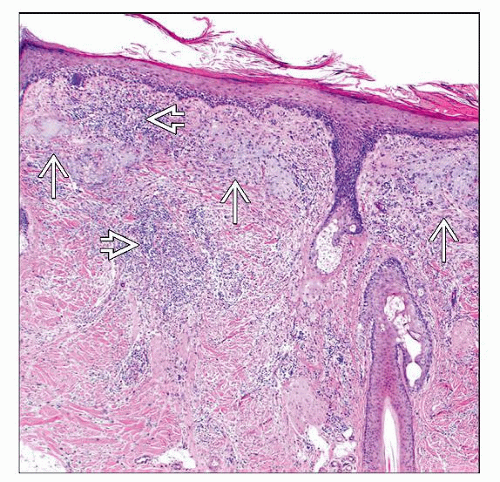 DM may resemble an inflamed scar on low-power examination. However, aggregates of lymphoid cells  and prominent solar elastosis and prominent solar elastosis  are usually identified and are helpful findings. are usually identified and are helpful findings. |
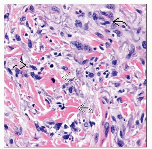 High-magnification examination shows hyperchromatic-staining atypical spindle cells  surrounded by abundant collagen surrounded by abundant collagen  bundles. bundles. |
TERMINOLOGY
Abbreviations
Desmoplastic melanoma (DM)
Synonyms
Desmoplastic/neurotropic melanoma
Definitions
Form of invasive melanoma composed of spindle cells associated with dense stromal collagen, resembling a scar
CLINICAL ISSUES
Epidemiology
Age
Presents in sun-damaged skin of elderly adults
Presentation
Firm skin-colored, tan or pink plaque or nodule
Sometimes depressed
Often amelanotic
Treatment
Surgical approaches
It is important to resect DM with clear surgical margins as early as possible for successful clinical management
There is increasing evidence that sentinel lymph node biopsy may not be indicated for “pure” variants of DM because of the low incidence of regional lymph node metastases
Prognosis
Tumor thickness
Clark level (IV vs. V)
Histological subtype: “Pure” (longer disease-free survival) vs. “combined”
“Pure” subtype is defined as > 90% scar-like areas
“Combined” subtype is defined as densely cellular spindle cell collections without significant scar-like areas (> 10%)
Tumor mitotic rate
MICROSCOPIC PATHOLOGY
Histologic Features
Ill-defined spindle cell neoplasm with highly infiltrative pattern of growth
Overlying epidermis may show melanoma in situ (usually lentigo maligna type)
Stromal collagen on scanning magnification resembles a scar
Spindle cells are arranged in fascicles and merge with scar-like areas
Cellular density and cytologic atypia can vary based on histologic subtype
Discrete dermal lymphoid aggregates
Solar elastosis


