Dermatofibrosarcoma Protuberans
David Cassarino, MD, PhD
Key Facts
Terminology
Low-grade malignant spindle cell tumor of skin characteristically showing prominent storiforming
Variant: Bednar tumor (pigmented DFSP)
Clinical Issues
Typically occurs in young adults
Excellent prognosis in most cases
Relatively low recurrence rate
Very low metastatic rates (usually only in cases with fibrosarcomatous transformation)
Microscopic Pathology
Dermal and subcutaneous involvement
Cells arrayed in storiform or cartwheel patterns
Proliferation of monomorphic spindle-shaped cells
Lesional cells lack significant pleomorphism
Mitoses usually infrequent (< 4/10 HPF)
Atypical mitoses usually absent
Ancillary Tests
CD34 is most reliable marker, typically strongly and diffusely positive
May be weak and focal in some cases
FXIIIA is typically negative
Focal staining, usually at periphery or in scattered dendritic cells
Top Differential Diagnoses
Cellular dermatofibroma/fibrous histiocytoma
Indeterminate fibrohistiocytic lesion
Fibrosarcoma (including transformation in DFSP)
Leiomyosarcoma
Spindle cell/desmoplastic melanoma
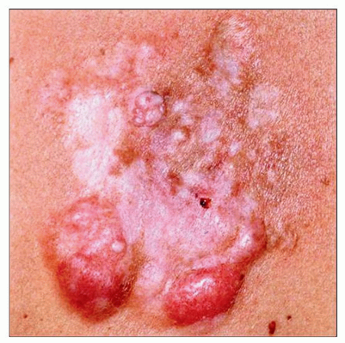 Dermatofibrosarcoma protuberans (DFSP) is often characterized clinically by an exophytic, multinodular growth with areas of interposed flattening or atrophy. |
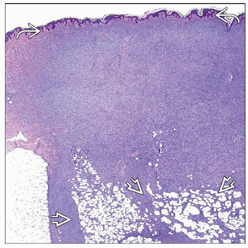 Low magnification of a DFSP shows deep dermal and subcutaneous  involvement by a cellular spindle cell tumor with fat entrapment. Epidermis is separated from tumor by thin grenz zone involvement by a cellular spindle cell tumor with fat entrapment. Epidermis is separated from tumor by thin grenz zone  . (Courtesy T. Mentzel, MD.) . (Courtesy T. Mentzel, MD.) |
TERMINOLOGY
Abbreviations
Dermatofibrosarcoma protuberans (DFSP)
Synonyms
Bednar tumor (pigmented DFSP)
Definitions
Low-grade malignant spindle cell tumor of skin characteristically showing prominent storiforming
ETIOLOGY/PATHOGENESIS
Unknown in Most Cases
Rare cases reportedly associated with previous trauma, burns, or arsenic exposure
Genetics
Rearrangements of collagen 1A1 (COL1A1)/plateletderived growth factor B (PDGFB)
Characteristic t(17;22) detected in most cases
Can be detected by FISH or PCR studies for the fusion protein
CLINICAL ISSUES
Epidemiology
Incidence
Uncommon tumors
Age
Typically occurs in young adults
Rare congenital cases reported
Gender
Male predominance
Site
Most often present on trunk or extremities
Rarely occur on head and neck
Presentation
Dermal and subcutaneous nodular/multinodular or plaque-like mass
Natural History
Slowly progressive, locally aggressive tumor
Treatment
Optimal treatment is complete surgical excision
Imatinib has been used for locally extensive and metastatic disease
Complete response reported in up to 50% of cases
Prognosis
Excellent in most cases
Local recurrences in up to 30% of cases
Very low metastatic potential (and essentially only in cases with fibrosarcomatous transformation)
MACROSCOPIC FEATURES
General Features
Polypoid, multinodular, or bosselated-appearing tumor
Rare cases may be atrophic appearing
Cut surface usually gray-white
May show hemorrhage and cystic changes
Size
Range: 1-10 cm
MICROSCOPIC PATHOLOGY
Histologic Features
Dermal and subcutaneous involvement
Proliferation of monomorphic spindle-shaped cells
Arrayed in storiform or cartwheel patterns
Lesional cells typically lack significant pleomorphism
Elongated spindle-shaped nuclei
Mild nuclear hyperchromasia, small to inconspicuous nucleoli
Moderate amounts of eosinophilic cytoplasm
Mitoses are usually infrequent (< 4/10 HPF) and not atypical
Increased mitoses and atypical forms are seen with fibrosarcomatous change
Necrosis is usually absent
Adnexal structures entrapped but not obliterated
Subcutaneous areas typically show “honeycombing” fat entrapment
Myxoid stromal change may be prominent in some cases
Cytologic Features
Elongated spindle-shaped cells with hyperchromatic-staining nuclei, small or absent nucleoli, and eosinophilic-staining cytoplasm
Variants
Bednar tumor
Pigmented DFSP due to intratumoral population of benign melanocytes
No prognostic significance
Giant cell fibroblastoma (GCFB)
Clinical: Occurs in children and young adults
Histologic features are distinctive
Proliferation of spindled cells and giant cells with nuclear hyperchromasia
Pseudovascular spaces lined by the giant cells
Mutations involving COL1A1/PDGFR (same as in DFSP)
ANCILLARY TESTS
Immunohistochemistry
Useful to confirm diagnosis, although often not necessary
CD34 is most reliable marker
Typically, strongly and diffusely positive
May be weak and focal in some cases
FXIIIA is usually negative (positive in dermatofibroma [DF])
May show focal staining, usually at periphery or in scattered dendritic cells
CD68, CD10, lysozyme, and chymotrypsin are typically negative
These markers are relatively nonspecific (but positive in DF and atypical fibroxanthoma)
Stay updated, free articles. Join our Telegram channel

Full access? Get Clinical Tree


