Chondroid Syringoma/Cutaneous Mixed Tumor Low Magnification Low magnification shows a well-circumscribed tumor composed of epithelial islands with easily identifiable glandular/ductal structures embedded in a myxoid and hyaline stroma.
Cutaneous Mixed Tumor With Ductal Lumina and Myxoid Stroma Small ducts lined by cuboidal cells are a common finding. The classic abundant myxoid and cartilaginous stroma is also seen .
Cutaneous Mixed Tumor at High Magnification Ductal structures with some foci of decapitation secretion are present in the epithelial component.
Cutaneous Mixed Tumor Composed of Basaloid Cells This chondroid syringoma is composed mostly of small, cuboidal-shaped basaloid cells surrounding multiple ductal lumina .
TERMINOLOGY
Abbreviations
• Cutaneous mixed tumor (CMT)
Synonyms
• Apocrine mixed tumor
• Eccrine mixed tumor
• Mixed tumor of folliculosebaceous-apocrine complex
Definitions
• Well-circumscribed benign epithelial tumor with chondromyxoid matrix
CLINICAL ISSUES
Epidemiology
• Incidence
Rare
• Age
Middle-aged to elderly
• Sex
Male predominance
• Ethnicity
No ethnic predilection
Site
• Head and neck sites are most common
• Extremities and trunk
Presentation
• Skin-colored, asymptomatic, dermal to subcutaneous tumors
Natural History
• Slow growth
Treatment
• Surgical excision
Prognosis
• Excellent
Very rare malignant transformation
MACROSCOPIC
Size
• 0.5-3.0 cm
Sections to Be Submitted
• Representative sections of grossly different-appearing areas should be submitted
MICROSCOPIC
Histologic Features
• Well-circumscribed dermal to subcutaneous tumor
• Composed of proliferation of epithelial cells embedded in myxoid, chondroid, or fibrous stroma
• Apocrine ducts within tumor can be seen
Many authors consider CMT to be predominately tumor of apocrine origin
• Differentiation toward different parts of folliculosebaceous-apocrine complex reported
Focal areas of matrical or sebaceous differentiation may rarely be seen
• Eccrine differentiation may be present in some cases
• Large amounts of adipose tissue are seen in what are called lipomatous mixed tumors
• Calcification and ossification are rare focal findings
• Due to shrinkage of stroma, fibroblasts and epithelial cells within stroma are surrounded by halos and appear similar to cells of cartilage
• Epithelial component
Epithelial cells can be arranged singly, in small and large clusters, or in solid cords
Ductal structures are of variable size and shape
CMTs have been divided into 2 types according to ductal structures by some
– Tumors with tubular, branching lumina
– Tumors with small, tubular lumina
Cystic dilation and branching of ducts is common
PAS(+), amorphous, eosinophilic material (collagenous spherulosis) can be seen in tubular lumina
Larger ducts lined by 2 layers of cuboidal cells and peripheral layer of myoepithelial cells
Some CMT composed predominantly of small ducts and small epithelial clusters
Small ducts lined by single layer of cuboidal cells
Only gold members can continue reading. Log In or Register to continue
 with easily identifiable glandular/ductal structures
  embedded in a myxoid and hyaline
  stroma.
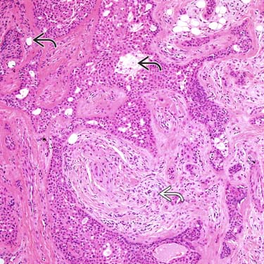
 lined by cuboidal cells are a common finding. The classic abundant myxoid and cartilaginous stroma is also seen
  .
.
 with some foci of decapitation secretion
  are present in the epithelial component.
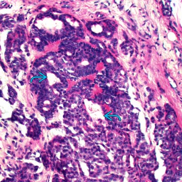
 .
.


