Cutaneous Angiosarcoma
Thomas Mentzel, MD
Key Facts
Terminology
Malignant mesenchymal neoplasm of cells variably recapitulating morphologic and functional features composed of endothelial cells
Etiology/Pathogenesis
Idiopathic (actinic) angiosarcoma
Predominantly in actinically damaged skin
Often initially mistaken for inflammatory lesion or cutaneous lymphoma/pseudolymphoma
Radiation-induced angiosarcoma
Angiosarcoma associated with chronic lymphedema
Clinical Issues
Needs wide excision with clear margins
Poor prognosis
Necrosis and epithelioid cytomorphology are adverse prognostic factors
Image Findings
Ill-defined, plaque- or bruise-like, often red lesions
Macroscopic Features
Ill-defined, hemorrhagic lesions
Microscopic Pathology
Vascular spaces of irregular size and shape
Anastomosing vascular channels
Multilayering, heaping up, and papillary formation of endothelial cells
Enlarged, hyperchromatic, irregular-shaped nuclei
No complete rim of actin(+) (myo)pericytes
Increased Ki-67 expression
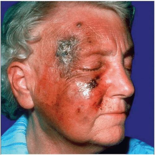 Clinical photograph shows an ill-defined, erythematous and plaque-like, hemorrhagic, cutaneous neoplasm in this male patient. |
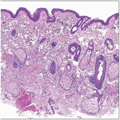 Hematoxylin & eosin shows a well-differentiated cutaneous angiosarcoma arising in sun-damaged skin. |
TERMINOLOGY
Abbreviations
Angiosarcoma (AS)
Synonyms
Hemangiosarcoma
Lymphangiosarcoma
Malignant hemangioendothelioma
Definitions
Malignant mesenchymal neoplasm of cells recapitulating variably morphologic and functional features composed of endothelial cells
Clear differentiation between lymphangiosarcoma and sarcoma with blood vessel differentiation remains problematic
ETIOLOGY/PATHOGENESIS
Developmental Anomaly
Congenital lymphedema
Environmental Exposure
Chronic lymphedema, i.e., after mastectomy (Stewart-Treves syndrome)
Radiotherapy
Sun exposure
CLINICAL ISSUES
Epidemiology
Incidence
Rare
Increasing rate due to use of radiotherapy and prolonged actinic damage
Age
More frequent in elderly patients
Very rare in children
Gender
M > F
Site
Relatively frequent in head and neck area
Radiation fields
Areas of acquired or congenital lymphedema
Presentation
Idiopathic (actinic) angiosarcoma
Arises predominantly in actinically damaged skin
Scalp, upper forehead, face
Elderly patients
Often initially mistaken for inflammatory lesion or cutaneous lymphoma/pseudolymphoma
Radiation-induced angiosarcoma
Arises at variable times after therapeutic irradiation (e.g., for breast cancer)
Angiosarcoma associated with chronic lymphedema
Congenital lymphedema
Acquired lymphedema (Stewart-Treves syndrome)
Ill-defined lesions
Plaque-like, red, indurated lesions
Bruise-like lesions
Nodular or multinodular appearance of older lesions
Often multifocal
Treatment
Surgical approaches
Wide excision with clear margins
Adjuvant therapy
Benefit of adjuvant/neoadjuvant chemotherapy unclear
IMAGE FINDINGS
General Features
Best diagnostic clue
Ill-defined, plaque-like lesions
Location
Actinically damaged skin of scalp and face
Specimen Radiographic Findings
Diagnostic pathologic vascular structures may be present
MACROSCOPIC FEATURES
General Features
Ill-defined, hemorrhagic lesions
Flat or ulcerated epidermis
Often sponge-like appearance
MICROSCOPIC PATHOLOGY
Histologic Features
Vascular spaces of irregular size and shape
Stay updated, free articles. Join our Telegram channel

Full access? Get Clinical Tree


