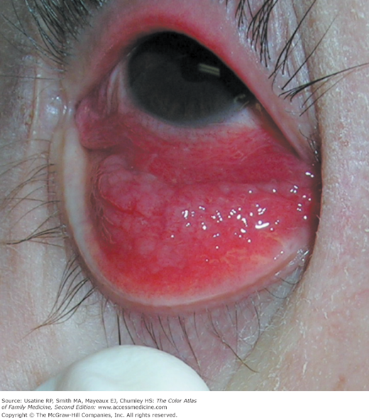
Patient Story
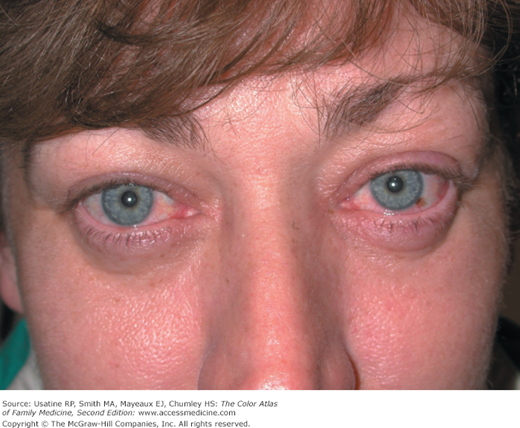
A 35-year-old woman presents with 2 days of redness and tearing in her eyes (Figure 16-1). She has some thin matter in eyes, but neither eye has been glued shut when she awakens. She does not have any trouble seeing once she blinks to clear any accumulated debris. Both eyes are uncomfortable and itchy, but she is not having any severe pain. She does not wear contact lenses and has not had this problem previously. The patient was diagnosed with viral conjunctivitis and scored −1 on the clinical scoring system (see “Diagnosis”). She was instructed about eye hygiene and recovered in 3 days.
Introduction
Conjunctivitis, inflammation of the membrane lining the eyelids and globe, presents with injected pink or red eye(s), eye discharge ranging from mild to purulent, eye discomfort or gritty sensation, and no vision loss. Conjunctivitis is most commonly infectious (viral or bacterial) or allergic, but can be caused by irritants. Diagnosis is clinical, based on differences in symptoms and signs.
Epidemiology
- Infectious conjunctivitis is common and often occurs in outbreaks, making the prevalence difficult to estimate.
- In the United States, the estimated annual incidence rate for bacterial conjunctivitis is 135 per 10,000 people.1
- Viral conjunctivitis is more common than bacterial conjunctivitis.
- Allergic conjunctivitis had a point prevalence of 6.4% and a lifetime prevalence of 40% in a large population study in the United States from 1988 to 1994.2
Etiology and Pathophysiology
Conjunctivitis is predominately infectious (bacterial or viral) or allergic, and the most common etiologies vary by age.
- Neonatal conjunctivitis is often caused by Chlamydia trachomatis and Neisseria gonorrhoeae.3
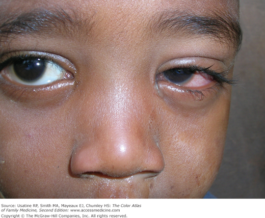
- Children younger than 6 years are more likely to have a bacterial than viral conjunctivitis (Figure 16-2). In the United States, the most common bacterial causes are Haemophilus species and Streptococcus pneumoniae accounting for almost 90% of cases in children.4
- Children age 6 years or older are more likely to have viral or allergic causes for conjunctivitis.4 Adenovirus is the most common viral cause.
Diagnosis
- To distinguish conjunctivitis from other causes of a red eye, ask about pain and check for vision loss. Patients with a red eye and intense pain or vision loss that does not clear with blinking are unlikely to have conjunctivitis and should undergo further evaluation.
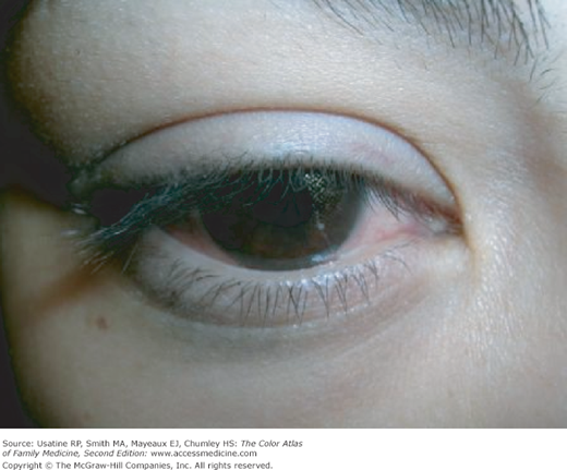
- Always ask about contact lens use as this can be a risk factor for all types of conjunctivitis, including bacterial conjunctivitis (Figure 16-3).
- Typical clinical features of any type of conjunctivitis may include eye discharge, gritty or uncomfortable feeling, one or both pink eyes, and no vision loss. The infection usually starts in one eye, and progresses to involve the other eye days later.
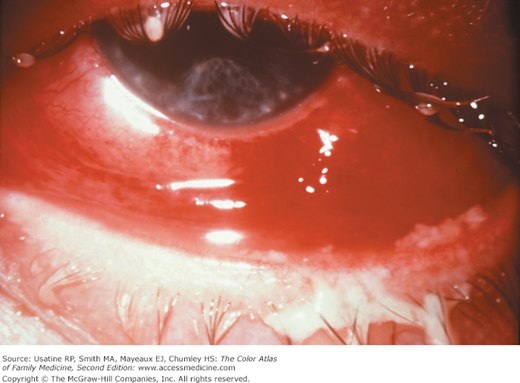
- Bacterial conjunctivitis (see Figures 16-2, 16-3, and 16-4) has a more purulent discharge than viral or allergic conjunctivitis.
- A clinical scoring system has been developed to distinguish bacterial from other causes of conjunctivitis in healthy adults who did not wear contact lenses. A score of +5 to −3 is determined as follows:
- Two glued eyes (+5); one glued eye (+2); history of conjunctivitis (−2); eye itching (−1).
- A score of +5, +4, or +3 is useful in ruling in bacterial conjunctivitis with specificities of 100%, 94%, and 92%, respectively.
- Scores of −1, −2, or −3 are useful in ruling out bacterial conjunctivitis with sensitivities of 98%, 98%, and 100%, respectively.5
- Two glued eyes (+5); one glued eye (+2); history of conjunctivitis (−2); eye itching (−1).
Allergic conjunctivitis is typically bilateral and accompanied by eye itching. Giant papillary conjunctivitis is a type of allergic reaction, most commonly to soft contact lenses (Figure 16-5).
- An in-office rapid test for adenoviral conjunctivitis (RPS Adeno Detector) has a sensitivity of 88% and a specificity of 91% compared to viral cell culture with confirmatory immunofluorescence staining.6
Stay updated, free articles. Join our Telegram channel

Full access? Get Clinical Tree


