Clear Cell Sarcoma
Elizabeth A. Montgomery, MD
Key Facts
Terminology
Sarcoma showing melanocytic differentiation
Malignant melanoma of soft parts
Clinical Issues
Young adults (3rd or 4th decade)
Usually affects extremities (> 90%)
Foot most common site
Behaves as high-grade sarcoma
Metastasizes to lymph nodes and lung
Often attached to tendon or aponeurosis
Occasional visceral examples
Ileum most common GI site
Microscopic Pathology
Monotonous clear to spindled cells arranged in nests/packets usually infiltrating tendon sheath
Prominent uniform nucleoli
Scattered wreath-like tumor giant cells
Most cases have few mitoses
Ancillary Tests
Immunohistochemical profile like that of melanoma in most cases
t(12;22)(q13;q12)
EWS-ATF1 fusion (detectable by PCR)
EWS-CREB1 fusion: Often in gastrointestinal CCS (lacks melanocytic differentiation)
Cases express S100 protein but not melanocytic markers
Same transcripts found in angiomatoid fibrous histiocytoma (different morphology, excellent prognosis)
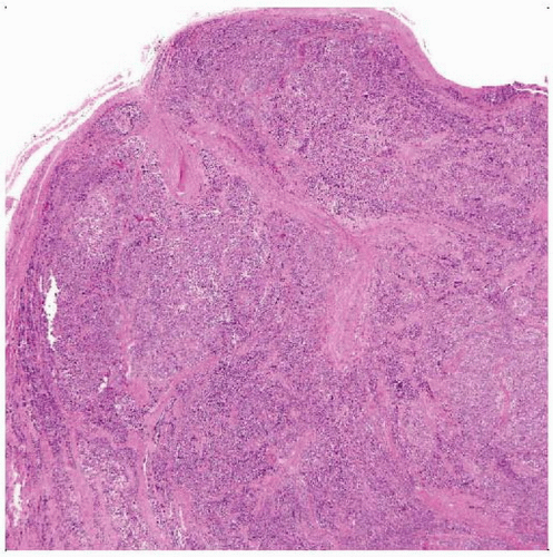
 Hematoxylin & eosin shows a lobulated (“packeted”) appearance of nests of neoplastic cells in a tendon or aponeurosis. |
TERMINOLOGY
Abbreviations
Clear cell sarcoma (CCS)
Synonyms
Malignant melanoma of soft parts
Definitions
Sarcoma showing melanocytic differentiation
Different from clear cell sarcoma of kidney
CLINICAL ISSUES
Epidemiology
Incidence
Rare
Age
Young adults (3rd or 4th decade)
Gender
Slight female predominance
Presentation
Painless mass
Usually affects extremities (> 90%)
Foot most common site
Often attached to tendon or aponeurosis
No associated skin lesions
Important in separating CCS from melanoma
Occasional visceral examples
GI tract most common visceral site
Ileum most common GI site
Prognosis
Behaves as high-grade sarcoma
5-year survival (50-65%)
Metastasizes to lymph nodes and lung
Differs from behavior of melanoma despite overlapping features
For example, 1 cm clear cell sarcoma may have good prognosis while 1 cm melanoma likely to be lethal
Metastatic pattern that of sarcoma
MACROSCOPIC FEATURES
Size
2-6 cm; median size: 2.5-3.5 cm
Lobulated gray-white masses
MICROSCOPIC PATHOLOGY
Histologic Features
Monotonous clear to spindled cells arranged in nests/packets usually infiltrating tendon sheath
Abundant glycogen can be detected by PAS or PAS with diastase
Prominent uniform nucleoli
Most cases lack necrosis
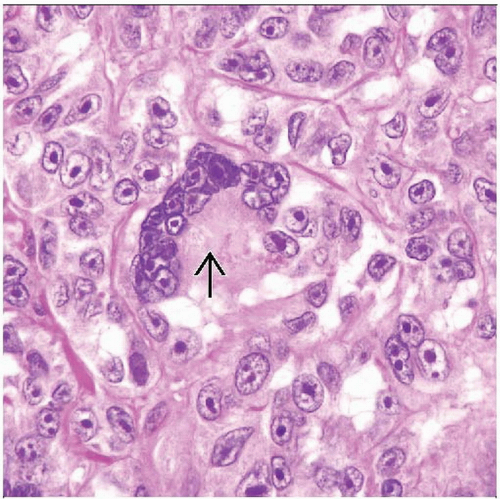
Scattered wreath-like tumor giant cells
Occasional melanin pigment
Rare cases have nuclear pleomorphism
Most cases have few mitoses
ANCILLARY TESTS
Immunohistochemistry
Profile like that of melanoma in most cases
Occasional synaptophysin, CD56, epithelial membrane antigen, cytokeratin AE1/AE3, CD34
Negative α-smooth muscle actin, desmin, and cytokeratin CAM5.2
Cytogenetics
t(12;22)(q13;q12)
Most soft tissue cases
t(2;22)(q13;q12)
Often in gastrointestinal cases
PCR
Useful for diagnosis in unusual cases
EWS-ATF1 fusion (detectable by PCR)
Found in 90% of soft tissue cases
Alternate EWS-CREB1 fusion
Same transcripts found in angiomatoid fibrous histiocytoma (which has different morphology and prognosis)
Noted in gastrointestinal CCS that lack melanocytic differentiation
Cases express S100 protein but not melanocytic markers
Melanocyte-specific splice form of MITF transcript found in examples with melanocytic differentiation
Gene Expression Profiling
Has overlap with cutaneous melanoma profile
DIFFERENTIAL DIAGNOSIS
Melanoma
Association with skin
Stay updated, free articles. Join our Telegram channel

Full access? Get Clinical Tree


