Chondroma
David R. Lucas, MD
Key Facts
Terminology
Benign hyaline cartilage neoplasm of soft tissue with predilection for hands and feet
Synonyms: Extraskeletal chondroma, chondroma of soft parts, osteochondroma, myxochondroma
Clinical Issues
Low recurrence rate (15-20%)
Recurrences controlled by reexcision
No reports of malignant degeneration
Image Findings
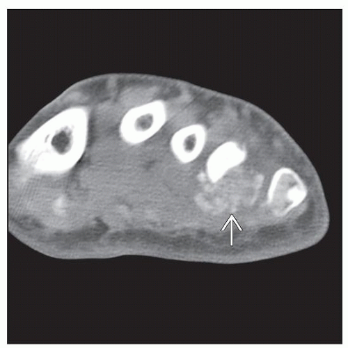
Small, well-demarcated, mineralized soft tissue mass in acral extremity
Macroscopic Features
Median size: 1.6 cm; range: 0.3-6.5 cm
Microscopic Pathology
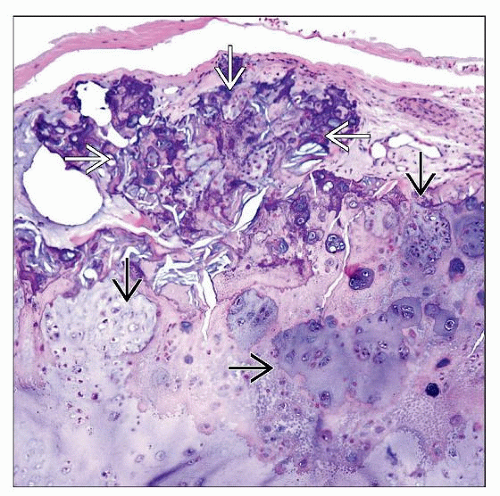
Well-circumscribed and lobulated
Mostly composed of mature hyaline cartilage
Variable amounts of calcification
Ossification common
Granulomatous inflammation in 15% of cases
Rare tumors with extensive xanthogranulomatous inflammation
Chondroblastoma-like chondroma
Top Differential Diagnoses
Tumoral calcinosis
Tophaceous pseudogout
Synovial chondromatosis
Extraskeletal myxoid chondrosarcoma
Calcifying aponeurotic fibroma
TERMINOLOGY
Synonyms
Extraskeletal chondroma, chondroma of soft parts, fibrochondroma, osteochondroma, myxochondroma, chondroblastoma-like chondroma of soft tissue
Definitions
Benign hyaline cartilage neoplasm of soft tissue with predilection for hands and feet
Only rare reports of primary cutaneous tumors (chondroma cutis)
CLINICAL ISSUES
Epidemiology
Incidence
Uncommon; exact incidence unknown
Exceedingly rare as cutaneous primary
Age
Median: 4th decade; range: Infancy to 9th decade
Gender
Women and men equally affected
Presentation
Painless mass
Most common in hands and feet (60-95%), especially fingers (40-50%)
Rare reports in proximal extremities, trunk, head and neck, upper aerodigestive tract, dura, skin, fallopian tube
Treatment
Surgical approaches
Simple excision
Prognosis
Low recurrence rate (15-20%)
Recurrences controlled by reexcision
No reports of malignant degeneration
IMAGE FINDINGS
General Features
Best diagnostic clue
Small, well-demarcated, mineralized soft tissue mass in acral extremity
Location
Hands and feet
Often in vicinity of joint or tendon
No intraarticular or subperiosteal localization by definition
Morphology
Most are calcified or ossified
Sometimes erode and deform underlying bone
MACROSCOPIC FEATURES
General Features
Well demarcated, spherical or ovoid
Stay updated, free articles. Join our Telegram channel

Full access? Get Clinical Tree


