Fig. 29.1
Xerosis: significant dryness of the skin of the foot
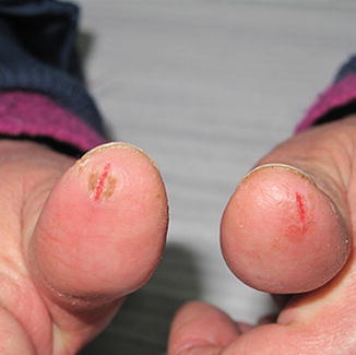
Fig. 29.2
Xerosis: fissures of the hands
Dry skin can be attributed to abnormal keratinocyte differentiation, which leads to a modification of stratum corneum and to an interference with sebaceous gland function, resulting into a loss of the water-retaining function of the epidermis. The development of chronic xerotic dermatitis can occur, exposing the skin to secondary infection with S. aureus and, rarely, herpes simplex virus.
Pharmacological Treatment
Short-term, low-dose topical steroids may be necessary for severe xerosis associated with inflammation. In case of infection with S. aureus, topical or systemic antibiotics should be used (clindamycin gel 1 % and flucloxacillin 500 mg three times daily for 7 days, or Doxycycline 100 mgs twice a day for 7 days). In cases of herpes simplex virus, antiviral drugs should be prescribed (valaciclovir 500 mg twice daily for 5 days).
Fissures can be treated with propylene glycol 50 % solution under plastic occlusion, salicylic acid 10 % ointment, hydrocolloid dressing, flurandrenolone tape, or liquid cyanoacrylate glue.
Dermocosmetological Treatment
Treatment of mild or moderate xerosis consists of thick moisturizing creams without fragrances or potential irritants. Specific creams can include urea, colloidal oatmeal, and petroleum-based creams. It is preferable to use unsaponifiable substance, aloe, niacinamide, tocopherols and tocotrienols, ceramides, gamma oryzanol, and cosmetic formulations with little or no presence of petrolatum and silicones, in order to avoid the maceration of the stratum corneum. Alcohol-containing lotions, retinoids, or benzoyl peroxide are not recommended. For scaly areas of xerosis, ammonium lactate or lactic acid creams can be utilized. For skin fissures, thick moisturizers or zinc oxide creams can be applied.
Patients should be instructed to avoid long-lasting baths and to use tepid water, to prefer detergents without surfactants and foaming substances, and bath oil or mild moisturizing soaps that are free from fragrances or perfumes.
Antimicrobial silk clothing can be an adjuvant strategy. This kind of fabric has extremely low frictional properties. The protein structure of fibroin is similar to stratum corneum of the human skin, able to absorb a high percentage of moisture without becoming damp, maintaining a stable heat, and constant skin temperature. It also has antimicrobial action, which could help to avoid secondary infections.
Alopecia
The overall incidence of chemotherapy-induced hair loss is estimated to be 65 %. The prevalence and severity are related to the chemotherapeutic agent: >80 % for anti-microtubule agents, 60–100 % for topoisomerase inhibitors, >60 % for alkylators, and 10–50 % for antimetabolites. Higher incidences are registered with poly-chemotherapy respect to monotherapy.
Chemotherapy-induced hair loss is considered to be one of the most traumatic aspects for oncological patients. It can negatively impact body image, sexuality, and self-esteem. 8 % of patients refuse to use chemotherapy for the anxiety of hair loss. Risk factors for hair loss are: drug dose, administration regimen, exposure to X-rays, age, comorbidities, the presence of androgenetic alopecia, and nutritional and hormonal status.
Hair-shaft shedding can occur days or weeks after the initiation of chemotherapy. Both shedding patterns, dystrophic anagen effluvium and telogen effluvium, can be observed. The affected areas seem to be selective, with a more frequent involvement of scalp regions that show low total hair densities, such as the frontal or occipital hairlines (Fig. 29.3). The mitotic activity of the hair follicle at the moment of the insult is one of the factors that may influence the shedding pattern.
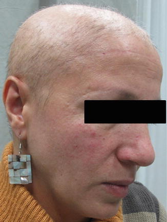
Fig. 29.3
Alopecia of the scalp in a patient treated with EGFRi
The highly proliferative matrix keratinocytes of anagen hair follicles, located in the hair bulb, and their pigmentary system are the main targets of chemotherapeutic drugs. They are very sensitive to toxins and drugs and can easily undergo rapid apoptosis. Sometimes, a damage of hair-follicle stem cells occurs; it can lead to permanent alopecia (Fig. 29.4).
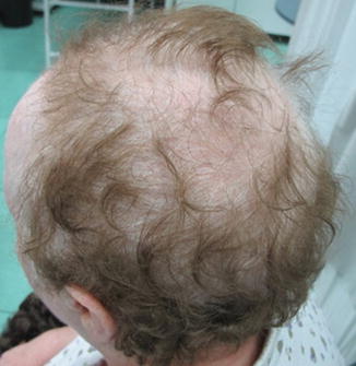
Fig. 29.4
Permanent alopecia in a patient treated with cyclophosphamide
Normally, up to 90 % of scalp hairs are in the anagen phase, for this reason the scalp is the most frequently affected area. Hairs of the beard, eyebrows, and eyelashes, as well as axillary and pubic regions, may be also affected, depending on the percentage of hairs in anagen. When hair is in late anagen phase, during which the mitotic rate is low, chemotherapy accelerates the normal transition to telogen, while catagen and telogen are not affected because they are mitotically inactive phases.
Generally, the hair loss is reversible, with hair regrowth typically occurring after a delay of 3–6 months. In some patients, the new growth shows changes in color and/or texture, but these differences can be temporary. Although rare, cases of permanent alopecia, in which hair regrowth is severely retarded or does not occur at all, are reported. This kind of alopecia is frequently associated with high-dose chemotherapy or with busulfan and cyclophosphamide administration.
Pharmacological Treatment
A solution of 2 % topical minoxidil is the best treatment for accelerating regrowth after chemotherapy. It must be applied twice a day on the involved areas for at least 1 month. Even if this type of alopecia is often reversible, it has been demonstrated that minoxidil reduces its severity and duration.
Dermocosmetological Treatment
A tetrapeptide composed of lysine, aspartic acid, valina, and tyrosine could be considered as an additional treatment. It is an analogue of P substance, a neuropeptide, which prolongs the anagen and retards the catagen phases, and acts as a releasing grow factor. It can be used in association with minoxidil, to improve its effect on hair regrowth, or it can also be used alone as maintenance treatment.
Implementation of gentle hair care strategies should be done not only throughout chemotherapy but also after. In order to avoid additional traumas, patients should use a soft brush, wash hair only as often as necessary, and use a gentle shampoo. Cutting hair short or shaving hair is not necessary, but it could be more comfortable.
A silk wig, glue-free, is the best way to make the patient feel better, avoiding additional irritation of the scalp (due to the glue and synthetic materials). This could help patients to deal with their condition and, at the same time, protect the scalp from sun and cold exposure.
Scalp cooling has been proposed as preventive therapy for alopecia. It is an effective method and patients’ compliance is good, except for some cases of headache and uncomfortable feelings reported. Some references indicate scalp cooling as a risk for scalp skin metastasis in hematological malignancies.
Papulo-Pustular Rash
A papulo-pustular rash is the most common cutaneous toxicity of EGFRi, affecting up to 90 % of patients. It is less frequent (30–40 %) and milder, with the MKi sorafenib and sunitinib.
The most frequently affected areas are sebaceous gland–bearing regions of the body, i.e., the scalp, face (Fig. 29.5), chest (Fig. 29.6), and upper aspect of the back, but the eruption can extend over the entire body except for the palms, soles, and mucosa. The rash commonly starts as a sensory disturbance characterized by erythema, edema, and dysesthesia (weeks 0–1), followed by a progression to inflammatory papules with central pustule formation (weeks 1–3), which result in the formation of crusts (weeks 3–5). The final phase is characterized by post-inflammatory pink or hyperpigmented macules and telangiectatic changes (weeks 5–8).
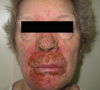
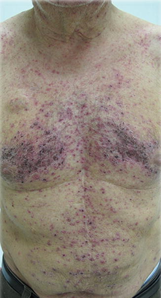
Fig. 29.5
Papulo-pustular rash of the face in a patient under treatment with panitumumab
Fig. 29.6
Papulo-pustular rash of the trunk in a patient under treatment with panitumumab
The pathogenesis of the acneiform eruption is not well understood. EGFR is expressed on epidermal keratinocytes, hair follicle epithelium, and the sweat gland apparatus. Its activation plays a crucial role in keratinocyte proliferation and differentiation, and in keratinization. Its inhibition induces growth arrest and apoptosis, decreasing cell migration, increasing cell attachment and differentiation, and stimulating inflammation.
Although the rash has been defined “acneiform” some differences have to be underlined. The EGFRI-induced papulopustular eruption does not present comedones, and differences in pathology and etiology from acne vulgaris exist. In acne, in fact, the primary process is sebaceous hyperplasia and lipid release into the follicular lumen; it leads to comedo formation and overgrowth of Propionibacterium acnes that results in follicular wall rupture, stimulating neutrophil chemotaxis and pustule formation. On the other hand, in EGFRi rash, the primary event is the damage to sebaceous glands and follicular epithelium, which leads to alteration in keratinocytes growth and differentiation. This causes the release of cytokines and the infiltration of mononuclear leucocytes (“sterile folliculitis”). The severity of the papulo-pustular rash is dose dependent and correlates with an improved tumor response and survival.
Even though it is never fatal, it has a negative impact on quality of life because of its visible characteristics and related symptoms, such as pain, burning, and skin sensitivity. Its main aggravating factors are sun exposure, concomitant radiotherapy, and inadequate moisture levels in the skin.
Pharmacological Treatment
The current guidelines for management of the EGFRi- associated papulo-pustular eruption include the following:
Grade 1 (mild): continue EGFRi; no treatment is required for rash; in some cases initiation of topical hydrocortisone 1 or 2.5 % cream and/or clindamycin 1 % gel could be necessary
Grade 2 (moderate): continue EGFRi; topical therapy plus systemic antibiotic therapy, such as doxycycline 100 mg twice daily, or oral minocycline 100 mg twice daily
Grade 3 (severe): reduce EGFRi dose; topical therapy plus oral antibiotic plus oral methylprednisolone from dose pack.
In uncontrolled trials, topical pimecrolimus and tacrolimus, and corticosteroids, showed clinical benefit. Advantages of choosing calcineurin inhibitors rather than corticosteroids include the absence of side effects such as skin fragility and the development of rosacea. Moreover, cyclosporine analogues could also theoretically partially reverse EGFR inhibition through their known ability to stimulate EGFR autophosphorylation, as shown in epidermoid cells, is well known. The potential role for retinoids (isotretinoin at low dose 20–30 mg/day) needs further investigation.
The beneficial effects of tetracyclines can be attributed to their anti-inflammatory and tissue-protective properties, through the inhibition of neutrophil and eosinophil chemotaxis; mitogen-induced lymphocyte proliferation; collagenases; and gelatinases.
Drugs used for the therapy of acne, including benzoyl peroxide and topical retinoids such as tretinoin, adapalene or tazarotene, are contraindicated because of irritation of the skin. Moreover, they haven’t shown clinical benefit in the treatment of the EGFRi rash.
Dermocosmetological Treatment
The patient should be instructed to:
use a thick alcohol-free highly occlusive moisturizing agent
favor tepid water and avoid prolonged, hot showers to minimize xerosis
use a broad-spectrum sunscreen
Non-occlusive make-up has been suggested to cover grade 1 and 2 rash, and it is well tolerated by patients. Recommended cosmetic formulations should contain fatty acids and ceramides, which are essential components of the stratum corneum barrier, and lactic acid that disrupts tightly adherent corneocytes, leading to the homogenization of the permeability barrier, although it should be used with caution because of the risk of irritation.
Nail Damage
Nail abnormalities are not uncommon during chemotherapy. The most frequent manifestations include Beau’s lines/onychomadesis, melanonychia, onycholysis (Fig. 29.7), and periungual pyogenic granulomas. It has been reported that taxanes cause nail changes more frequently than other drugs.
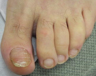
Fig. 29.7
Taxane-induced onycholysis
Some nail abnormalities, such as dark pigmentations, Mees’ lines and Beau’s lines, are asymptomatic. After the end of chemotherapy, they migrate distally as the nail grows, and usually no new stripes develop. However, other changes such as sub-ungual hemorrhage, paronychia, and onycholysis (loss of the nail plate) can negatively affect the quality of life of the patient, causing pain and impairment to activities of daily life.
Paronychia and periungual pyogenic granuloma-like lesions are observed in 10–30 % of patients receiving EGFRi, developing after 2–3 months after drug exposure (Fig. 29.8). Paronychia is characterized by edematous inflammation of the nail folds and usually affects the first digit. Periungual pyogenic granuloma-like lesions are characterized by easily bleeding, friable vascular tissue overgrowth on lateral nail folds.
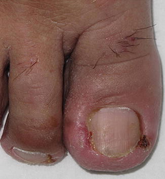
Fig. 29.8
Lapatinib-induced paronychia of the great toe
The pathogenesis of paronychia could be explained by the presence of a traumatic conflict between the thin tissues around the nail and nail itself. In fact, changes in growth and differentiation of the nail are responsible for the retention of squamous epithelium in the nail folds, which act as foreign bodies, causing an inflammatory reaction.
Pharmacological Treatment
Treatment of paronychia can be difficult, consisting of symptomatic relief with soaks, such as aluminum acetate or Burow’s solution, and cushioning of the affected areas, in addition to treatment with topical or systemic antibiotics. For granulomas, the use of silver nitrate sticks can also be useful. In severe cases, the affected nail may need to be removed.
A new proposal treatment is represented by 8 % phenol cauterization. It has the advantages to be a conservative, effective, simple approach that can be performed without a previous anesthesia. These aspects increase the compliance with the therapy. Cessation of therapy with EGFRi may be required in severe cases to allow healing.
Dermocosmetological Treatment
The patient should be instructed to avoid tight shoes, frequent water immersion, and contact with chemicals. As for xerosis, a useful adjuvant instrument is antimicrobial silk gloves and soaks.
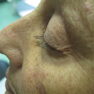
Trichomegaly
Trichomegaly of the eyelashes is characterized by a paradoxical overgrowth of eyelashes. It is a rare adverse effect of EGFRi, which usually occurs 2–5 months after the start of treatment and may resolve in several weeks or months after its discontinuation. The pathogenesis is not completely understood. EGFR is expressed in the keratinocytes of the outer sheath of the hair follicle. Its inhibition arrests the progression from anagen to telogen phase, leading to an aberrant anagen phase and, subsequently, to abnormal hair growth and to the formation of a disorganized hair follicle (Figs. 29.9 and 29.10).