54
Cestodes
CHAPTER CONTENTS
INTRODUCTION
Platyhelminthes (platy means flat; helminth means worm) are divided into two classes: Cestoda (tapeworms) and Trematoda (flukes). The trematodes are described in Chapter 55.
Tapeworms consist of two main parts: a rounded head called a scolex and a flat body consisting of multiple segments. Each segment is called a proglottid. The scolex has specialized means of attaching to the intestinal wall, namely, suckers, hooks, or sucking grooves. The worm grows by adding new proglottids from its germinal center next to the scolex. The oldest proglottids at the distal end are gravid and produce many eggs, which are excreted in the feces and transmitted to various intermediate hosts such as cattle, pigs, and fish.
Humans usually acquire the infection when undercooked meat or fish containing the larvae is ingested. However, in two important human diseases, cysticercosis and hydatid disease, it is the eggs that are ingested and the resulting larvae cause the disease.
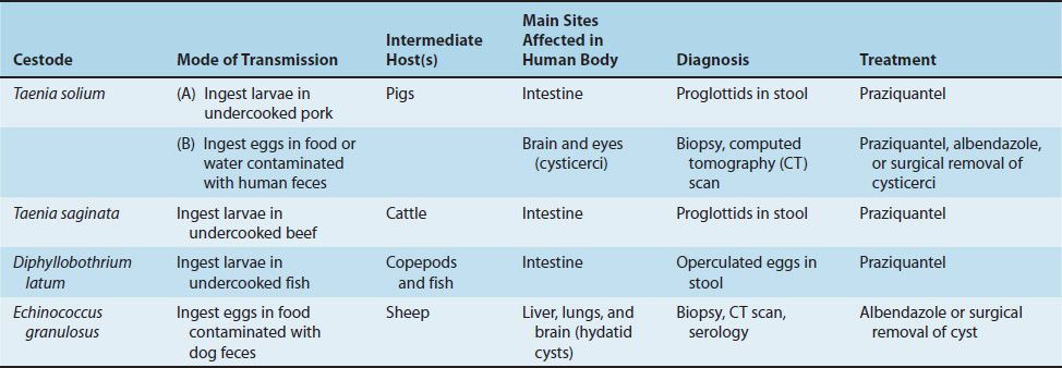
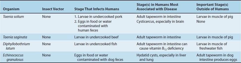
There are four medically important cestodes: Taenia solium, Taenia saginata, Diphyllobothrium latum, and Echinococcus granulosus. Their features are summarized in Table 54–1, and the medically important stages in the life cycle of these organisms are described in Table 54–2. Three cestodes of lesser importance, Echinococcus multilocularis, Hymenolepis nana, and Dipylidium caninum, are described at the end of this chapter.
TAENIA
There are two important human pathogens in the genus Taenia: T. solium (the pork tapeworm) and T. saginata (the beef tapeworm).
1. Taenia solium
Disease
The adult form of T. solium causes taeniasis. T. solium larvae cause cysticercosis.
Important Properties
The life cycle of T. solium is shown in Figure 54–1. T. solium can be identified by its scolex, which has four suckers and circle of hooks, and by its gravid proglottids, which have 5 to 10 primary uterine branches (Figures 54–2A, B and 54–3). The eggs appear the same microscopically as those of T. saginata and Echinococcus species (Figure 54–4A).
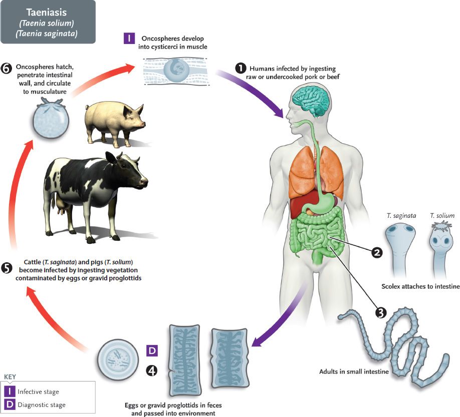
FIGURE 54–1 Taenia solium and Taenia saginata. Life cycle. Right side of figure describes the stages within the human (blue arrows). Humans are infected at step 1 when they ingest undercooked pork (T. solium) or beef (T. saginata) containing cysticerci (larval stage). Adult tapeworms form in intestine and lay eggs. Pigs and cattle are infected when they ingest either the eggs or proglottids in human stool. Left side of figure describes the stages within the pigs and cattle (red arrows). (Provider: Centers for Disease Control and Prevention/Dr. Alexander J. da Silva and Melanie Moser.)
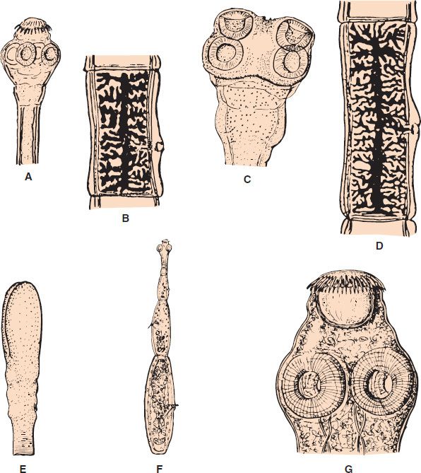
FIGURE 54–2 A: Taenia solium scolex with suckers and hooks (10×). B: Taenia solium gravid proglottid. This has fewer uterine branches than does the proglottid of Taenia saginata (see panel D) (2×). C: T. saginata scolex with suckers (10×). D: T. saginata gravid proglottid (2×). E: Diphyllobothrium latum scolex with sucking grooves (7×). F: Entire adult worm of Echinococcus granulosus (7×). G: E. granulosus adult scolex (70×).
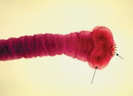
FIGURE 54–3 Taenia solium—scolex and several proglottids. Long arrow points to one of the four suckers on the scolex of T. solium. Short arrow points to the circle of hooklets. Proglottids can be seen extending from the scolex toward the left side of the image. (Figure courtesy of Dr. M. Melvin, Public Health Image Library, Centers for Disease Control and Prevention.)
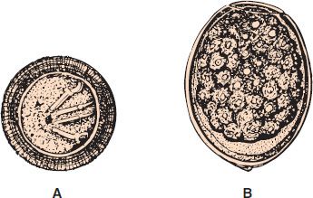
FIGURE 54–4 A: Taenia solium egg containing oncosphere embryo. Four hooklets are visible. Taenia saginata and Echinococcus granulosus eggs are very similar to the T. solium egg but do not have hooklets. B: Diphyllobothrium latum egg with an operculum on the top (300×).
In taeniasis, the adult tapeworm is located in the human intestine (Figure 54–1). This occurs when humans are infected by eating raw or undercooked pork containing the larvae, called cysticerci. (A cysticercus consists of a pea-sized fluid-filled bladder with an invaginated scolex.) In the small intestine, the larvae attach to the gut wall and take about 3 months to grow into adult worms measuring up to 5 m. The gravid terminal proglottids containing many eggs detach daily, are passed in the feces, and are accidentally eaten by pigs. Note that pigs are infected by the worm eggs; therefore, it is the larvae (cysticerci) that are found in the pig. A six-hooked embryo (oncosphere) emerges from each egg in the pig’s intestine. The embryos burrow into a blood vessel and are carried to skeletal muscle. They develop into cysticerci in the muscle, where they remain until eaten by a human. Humans are the definitive hosts, and pigs are the intermediate hosts.
In cysticercosis, a more dangerous sequence occurs when a person ingests the worm eggs in food or water that has been contaminated with human feces (Figure 54–5). Note that in cysticercosis, humans are infected by eggs excreted in human feces, not by ingesting undercooked pork. Also, pigs do not have the adult worm in their intestine, so they are not the source of the eggs that cause human cysticercosis. The eggs hatch in the small intestine, and the oncospheres burrow through the wall into a blood vessel. They can disseminate to many organs, especially the eyes and brain, where they encyst to form cysticerci (Figure 54–6). Each cysticercus contains a larva.
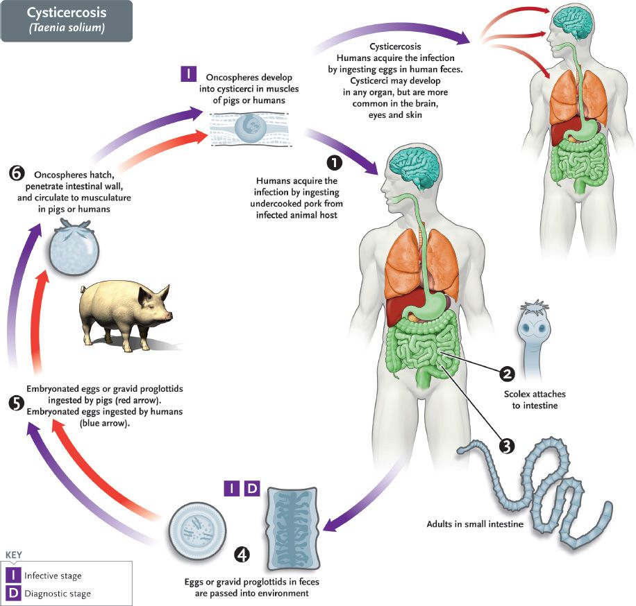
FIGURE 54–5 Taenia solium. Life cycle including cysticercosis stage. Center and left side of figure describes the cycle of T. solium within the human and the pig similar to Figure 54–1. Note, however, that there are now blue arrows between the egg at the bottom that go up the left side of the figure to the person at the top right. In cysticercosis, humans are infected when they ingest the eggs of T. solium in food contaminated with human feces. The eggs differentiate into cysticerci primarily in brain, eyes, and skin. (Provider: Centers for Disease Control and Prevention/Dr. Alexander J. da Silva and Melanie Moser.)
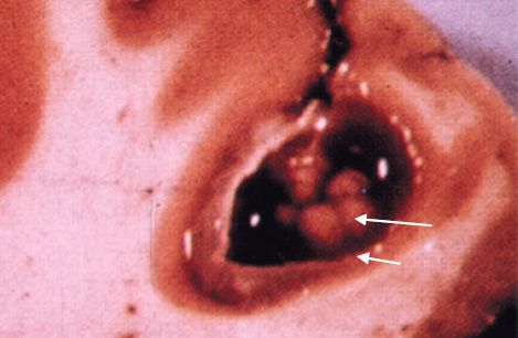
FIGURE 54–6 Cysticercus of Taenia solium in brain—long arrow points to a larva of T. solium. Short arrow points to the wall of the cysticercus (sac) that surrounds the larva. (Figure courtesy of Rhodes B. Holliman, PhD, Professor Emeritus, Virgina Tech.)
Pathogenesis & Epidemiology
The adult tapeworm attached to the intestinal wall causes little damage. The cysticerci, on the other hand, can become very large, especially in the brain, where they manifest as a space-occupying lesion (Figure 54–6). Living cysticerci do not cause inflammation, but when they die, they can release substances that provoke an inflammatory response. Eventually, the cysticerci calcify.
The epidemiology of taeniasis and cysticercosis is related to the access of pigs to human feces and to consumption of raw or undercooked pork. The disease occurs worldwide but is endemic in areas of Asia, South America, and Eastern Europe. Most cases in the United States are imported.
Clinical Findings
Most patients with adult tapeworms are asymptomatic, but anorexia and diarrhea can occur. Some may notice proglottids in the stools. Cysticercosis in the brain causes headache, vomiting, and seizures. Cysticercosis in the eyes can appear as uveitis or retinitis, or the larvae can be visualized floating in the vitreous. Subcutaneous nodules containing cysticerci commonly occur. Cysts also are commonly found in skeletal muscle.
Laboratory Diagnosis
Identification of T. solium consists of finding gravid proglottids with 5 to 10 primary uterine branches in the stools. In contrast, T. saginata proglottids have 15 to 20 primary uterine branches. Eggs are found in the stools less often than are proglottids. Diagnosis of cysticercosis depends on demonstrating the presence of the cyst in tissue, usually by surgical removal or computed tomography (CT) scan. Serologic tests (e.g., enzyme-linked immunosorbent assay [ELISA]) that detect antibodies to T. solium antigens are available, but they may be negative in neurocysticercosis.
Treatment
The treatment of choice for the intestinal worms is praziquantel. The treatment for cysticercosis is either praziquantel or albendazole, but surgical excision may be necessary.
Prevention
Prevention of taeniasis involves cooking pork adequately and disposing waste properly so that pigs cannot ingest human feces. Prevention of cysticercosis consists of treatment of patients to prevent autoinfection plus observation of proper hygiene, including handwashing, to prevent contamination of food with the eggs.
2. Taenia saginata
Disease
T. saginata causes taeniasis. T. saginata larvae do not cause cysticercosis.
Important Properties
T. saginata has a scolex with four suckers but, in contrast to T. solium, no hooklets. Its gravid proglottids have 15 to 25 primary uterine branches, in contrast to T. solium proglottids, which have 5 to 10 (Figure 54–1C and D). The eggs are morphologically indistinguishable from those of T. solium.
The life cycle of Taenia saginata is shown in Figure 54–1. Humans are infected by eating raw or undercooked beef containing larvae (cysticerci). In the small intestine, the larvae attach to the gut wall and take about 3 months to grow into adult worms measuring up to 10 m (Figure 54–7). The gravid proglottids detach, are passed in the feces, and are eaten by cattle. The embryos (oncospheres) emerge from the eggs in the cow’s intestine and burrow into a blood vessel, where they are carried to skeletal muscle. In the muscle, they develop into cysticerci. The cycle is completed when the cysticerci are ingested. Humans are the definitive hosts and cattle the intermediate hosts. Unlike T. solium, T. saginata does not cause cysticercosis in humans.
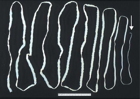
FIGURE 54–7 Taenia saginata—adult tapeworm. Note the tiny scolex on the right side of the image and the gravid proglottids on the left side of the image. White arrow points to the scolex. Ruler is 12 inches long. (Figure courtesy of Public Health Image Library, Centers for Disease Control and Prevention.)
Stay updated, free articles. Join our Telegram channel

Full access? Get Clinical Tree


