Clear cells show small, round to oval monomorphous nuclei; eosinophilic or clear cytoplasm
Heavily pigmented spindled and dendritic cells alternate with clear cells
Top Differential Diagnoses
• Atypical cellular blue nevus
• Melanoma arising in or mimicking cellular blue nevus (malignant blue nevus)
• Desmoplastic melanoma
Diagnostic Checklist
• Clinical features
Heavily pigmented black or blue nodule or plaque
• Pathologic features
Should not show multiple mitoses or necrosis (both of which favor malignancy)
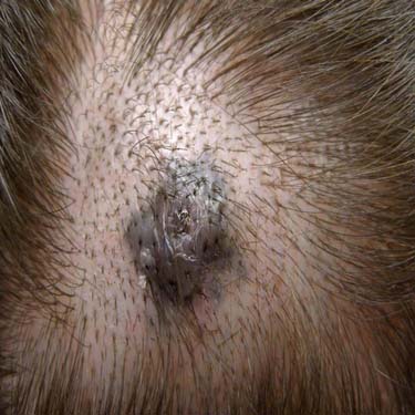
Clinical Image of a CBN Gray to dark blue tumor on the scalp of a middle-aged woman shows central ulceration related to trauma. (Courtesy J. Finch, MD.)
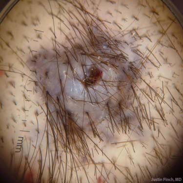
Dermatoscopic Image of a CBN Dermoscopy of the same lesion shows a uniformly gray nodule. (Courtesy J. Finch, MD.)
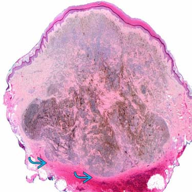
CBN at Scanning Magnification At this low power, a conventional blue nevus (CBN) showing nodular growth with deep pushing border is identified.
CBN at Low Magnification Showing Base of Lesion The lesion is heavily pigmented and shows small nests/nodules , of oval to spindle-shaped melanocytes.
TERMINOLOGY
Abbreviations
• Cellular blue nevus (CBN)
Definitions
• Uncommon cellular variant of blue nevus
• Presents as large, blue to blue-black, well-circumscribed, multilobulated tumor composed of oval to spindle-shaped melanocytes
CLINICAL ISSUES
Epidemiology
• Age
Occurs in childhood and young adult life; mean: 33 years
• Sex
Female predominance (F:M ~ 2:1)
• Ethnicity
All affected
Site
• Mostly occurs on sacrococcygeal regions and buttock but may also be located on scalp, neck, face, hands, and extremities (especially dorsal foot)
Presentation
• Heavily pigmented, slow-growing, black or blue nodule or plaque typically ranging 1-2 cm and sometimes up to 6 cm in diameter
• Ulceration may be seen in lesions appearing in area with growth restriction, such as dorsal foot
• Rarely can be with satellitosis, characterized clinically by development of macules around a central papule or nodule, and may thus mimic melanoma
Treatment
• Surgical approaches
Simple excision
Prognosis
• Benign but may rarely recur
• Regional lymph node involvement and benign metastases to regional lymph nodes with CBN are rare, but well documented
Resulting lymphadenopathy is prone to misdiagnosis as metastatic malignant melanoma
• Has rare potential for malignant transformation, and affected patients have poor clinical outcome
MICROSCOPIC
Histologic Features
• Architecture consists of central mass with adjacent ramifications
These have dumbbell or peninsula-like shape occupying place of effaced hair follicles, deeply extending into subcutis
Only gold members can continue reading. Log In or Register to continue


 is identified.
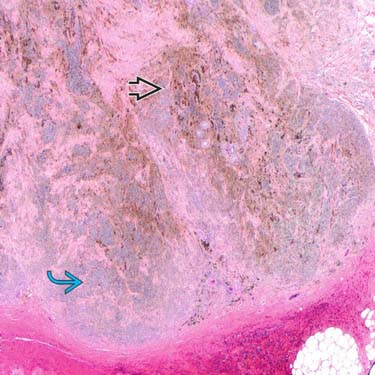
 and shows small nests/nodules
  , of oval to spindle-shaped melanocytes.


 These have dumbbell or peninsula-like shape occupying place of effaced hair follicles, deeply extending into subcutis


