• Numbness, tingling, and/or burning pain on palmar surfaces of first three digits and lateral half of fourth digit of the hand. • Positive Tinel’s sign (tingling or shocklike pain on volar wrist percussion). • Positive Phalen’s sign (appearance or exacerbation of symptoms caused by flexion of wrist for 60 seconds). • Electromyography may show decreased nerve conduction velocity across carpal tunnel. • Symptoms: pain and tingling or numbness in hand; worse at night and may awaken patient; relief from shaking or rubbing hand; can be bilateral; clumsiness from inability to hold or feel an object. • Physical examination: impaired sensation in distribution of median nerve. If condition is prolonged, atrophy of thenar eminence and weakness of thumb abductor may be present. Tinel’s and Phalen’s tests are accepted for diagnosing CTS in general practice. Tinel’s sign is tingling in the distribution of the median nerve when tapping nerve in middle of palm over the transverse carpal ligament. Phalen’s test is positive when symptoms are reproduced by holding the wrist in forced flexion for 60-90 seconds. Katz and Stirrat devised a self-administered hand diagram that is 80% sensitive and 90% specific for classic or probable CTS. Another in-office test is subjective assessment of swelling in the hand and palpation of the hand and wrist. Continued subjective swelling during treatment with wrist splinting correlates with poorer clinical response. Palpation of wrist and thumb in various directions of motion is a predictor of CTS. Wrist motion restriction correlates with electromyographic (EMG) diagnosis of CTS. • Other diagnostic tests: EMG helps confirm median nerve compression in the carpal tunnel. Nerve conduction velocity is normal from elbow to wrist and diminished from wrist to hand and fingers. Both sensory and motor nerve conduction velocities are measured. Magnetic resonance imaging and high-definition ultrasound imaging measure median nerve size and interior dimensions of the carpal tunnel. These tests are used for preoperative assessment of CTS to choose the best approach to surgery. • Differential diagnosis: CTS paresthesia is confused with paresthesia of brachial plexus in thoracic outlet syndrome or paresthesia of radial or ulnar nerves. Hand pain may be presenting feature of reflex sympathetic dystrophy (shoulder-hand syndrome). Also consider impingement of median nerve below the elbow, where the nerve passes under the pronator teres muscle. Specific dermatome pattern of CTS, without proximal dysfunction, should lead to history and tests establishing diagnosis of CTS. Left-sided CTS may be confused with angina pectoris.
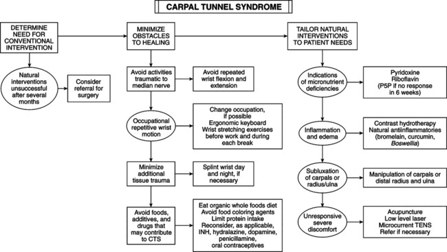
Carpal Tunnel Syndrome
DIAGNOSTIC SUMMARY
DIAGNOSTIC CONSIDERATIONS
![]()
Stay updated, free articles. Join our Telegram channel

Full access? Get Clinical Tree


Basicmedical Key
Fastest Basicmedical Insight Engine

