Introduction
Diagnostic accuracy when assessing patients with cardiovascular disease relies heavily on the medical history. Many patients with ischaemic heart disease have few or no physical signs and a characteristic history of peripheral vascular disease may be elicited. Key features in the cardiovascular history are shown in Box 3.1.
 Box 3.1 Important Features in the Cardiovascular History
| Chest pain: | onset |
| duration | |
| nature | |
| precipitating factors | |
| relieving factors | |
| distribution and radiation | |
| previous episodes | |
| associated symptoms (nausea, vomiting, pallor) | |
| Breathlessness: | on exertion |
| orthopnoea | |
| paroxysmal nocturnal dyspnoea | |
| Cough: | with or without sputum production |
| Oedema: | ankle swelling |
| swelling of lower limbs and sacral area | |
| Syncope: | on exertion |
| postural | |
| sudden | |
| Calf pain: | intermittent claudication |
| Peripheral vascular disease (PVD): | cold peripheries with colour/sensory changes |
| Others: | transient hemiparesis |
| Visual disturbance: | transient (e.g. amaurosis fugax) |
| Risk factors: | smoking |
| obesity | |
| hypertension | |
| diabetes | |
| hyperlipidaemia | |
| oral contraception | |
| Past history: | hypertension |
| cerebrovascular disease | |
| peripheral vascular disease | |
| congenital heart disease | |
| Medication: | antihypertensives |
| antianginal therapy | |
| statins | |
| oral contraceptives and estrogen replacement therapy (HRT) | |
| Family history: | ischaemic heart disease |
| diabetes | |
| hypertension | |
| hyperlipidaemia |
Systematic and thorough examination of the cardiovascular system is a core skill for physicians. Accurate assessment of peripheral cardiovascular signs aids the interpretation of auscultatory findings. Patients with ischaemic heart disease may have few physical signs and physicians should be aware of the likely sites and significance of scars from previous surgical or radiological intervention. Cardiac valvular disease and septal defects usually give rise to murmurs which may be diagnostic. In clinical practice arrival at the final cardiac diagnosis is aided by an electrocardiogram (ECG), chest X-ray (CXR) and echocardiogram (ECHO) and by more complex radiological intervention as appropriate including magnetic resonance imaging (MRI), computerised tomography (CT) and angiography.
General Inspection
Note
- peripheral or central cyanosis: central cyanosis is accompanied by peripheral cyanosis by definition
- dyspnoea and orthopnoea
- malar flush
- xanthelasmata
Blood Pressure
- measure the blood pressure lying and standing
Hands
Inspect for
- clubbing
- splinter haemorrhages
- palmar erythema
- nicotine staining
Arterial Pulses
Palpate
- radial pulse to assess the rate and rhythm
- radial and brachial pulses in both arms, comparing right and left
- carotid pulses
- radial and femoral pulses simultaneously to assess radiofemoral delay
Rate
- count radial for at least 15 s if rhythm regular, at least 30–60 s if irregular
- check the jugular venous pressure (JVP) whilst counting
Rhythm
- regular (sinus rhythm)
- regularly irregular (extra or dropped beats)
- irregularly irregular (atrial fibrillation; the pulse rate is different from the heart rate; listen and count at the apex whilst palpating the pulse)
Volume and character (Table 3.1)
- collapsing pulse: raise the patient’s arm above the level of the heart whilst holding four fingers over the anterior forearm
- slow – rising, low volume, alternans, bisferiens and paradoxus by palpating the carotid pulse
Table 3.1 Abnormalities of the Arterial Pulse
| Type | Character | Seen in |
| Slow-rising | Low amplitude, slow rise, slow fall | Aortic stenosis |
| Collapsing | Large amplitude, rapid rise, rapid fall | Aortic incompetence; severe anaemia, hyperthyroidism; arteriovenous shunt, heart block, patent ductus arteriosus |
| Low volume | Thready | Low cardiac output states; hypovolaemic shock; valvular stenosis; pulmonary hypertension |
| Alternans | Alternate large- and small-amplitude beats (rarely noted in pulse; usually on taking blood pressure) | Left ventricular failure |
| Bisferiens | Double-topped (‘notched’) | Aortic stenosis with aortic incompetence |
| Paradoxus | Pulse volume decreases excessively with inspiration | Cardiac tamponade, constrictive pericarditis, severe inspiratory airways obstruction |
| Absent radial | Congenital anomaly (check brachials and blood pressure) | |
| Tied off at surgery or catheterisation | ||
| Arterial embolism |
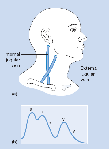
Jugular Venous Pressure and Pulse (Table 3.2; Fig. 3.1)
The patient should be lying at 45° with the neck relaxed. The JVP is seen welling up between the two heads of sternomastoid in the front of the neck on expiration.
Table 3.2 Raised Jugular Venous Pressure (JVP)
| Character | Compression of neck and abdomen | Conclusion |
| Non-pulsatile | No change in JVP | Superior mediastinal obstruction |
| • carcinoma of bronchus | ||
| • large goitre | ||
| • platysmal compression | ||
| Pulsatile | Jugular vein fills and empties | Right heart failure |
| Expiratory airways obstruction | ||
| Fluid overload | ||
| Cardiac tamponade |
Figure 3.1 (a) Surface markings of the internal and external jugular veins. (b) Jugular venous pulse waveform.

Stay updated, free articles. Join our Telegram channel

Full access? Get Clinical Tree


