Body Fluids and Water Balance


Body Fluids and Water Balance


Hwai-Ping Sheng, PhD


Physiological Functions of Water


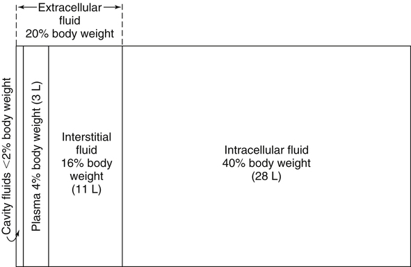
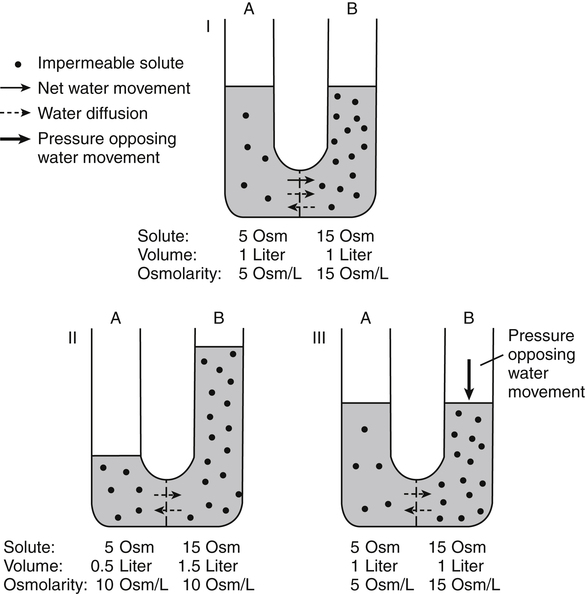
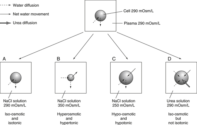
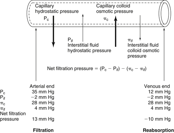
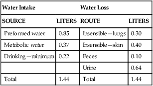
Water is an essential nutrient vital to the existence of both animals and plants. In the body, water is present inside and around the cells and within all blood vessels. It lubricates joints and moistens tissues such as those in the eyes, nose, and mouth. The volume of the intracellular fluid provides turgor to the tissues, which is important for the tissue or organ form and ultimately the body form. Water performs several functions that are essential to life. It is the principal fluid medium in which nutrients, minerals, gases, and enzymes are dissolved. The extracellular water bathing the cells serves as a medium for the transport of nutrients and oxygen to the cells and for removing wastes from the cells, which will be eliminated by the liver and kidneys. The intracellular water establishes the physicochemical medium that allows various metabolic processes to take place. Another important physiological function of water is its role in the regulation of body temperature. This is achieved by removing excess heat from the body by evaporative water loss from the skin.


Body Water Compartments


Body Water Content


Water makes up the largest component of the body; its content in the body varies with age, sex, and adiposity of the individual. In the neonate, water makes up approximately 75% of body weight, decreasing progressively to about 60% in the young adult, and continuing to decline to approximately 50% at about 50 years of age. The higher proportion of water in the neonate is for the most part the result of a larger fraction of its body mass as extracellular fluid space. A combination of factors causes the proportion of extracellular fluid space to decrease gradually with an increase in age. These factors include an increase in the amount of cellular tissues, such as muscles, at the expense of extracellular space and an increase in the proportion of body mass made up of adipose tissues and the supporting structures of skeleton, cartilage, and connective tissues, all of which contain a relatively low water content.


Adult women have lower water content when compared with men of comparable age, and obese individuals have lower water content than their leaner counterparts. These variations can be attributed to differences in the proportion of adipose tissue relative to lean tissue in the body. Fat cells have a relatively low content of water, about 10%, whereas other cellular tissues such as muscles contain an average of 70% water. Therefore water content in the body varies inversely with the relative proportion of adipose tissue, and this can explain the lower water content both in women and in obese individuals. However, when body water is calculated on a lean body weight basis, it constitutes a relatively constant proportion, 73.2% for adults and 82% for neonates.


Distribution of Body Water


Water in the body is distributed throughout the various body fluid compartments. The simplest subdivision is into an intracellular and an extracellular compartment, with the two compartments separated by the cell membrane (Figure 35-1). On average, a 70-kg adult has 42 L of water, of which two thirds (28 L) is intracellular and one third (14 L) is extracellular. The extracellular fluid is made up of the interstitial fluid (11 L), which bathes the cells and includes the lymph, the plasma (3 L), and the cavity or transcellular fluids, of which the largest volume is secretory fluids in the lumen of the gastrointestinal tract.


The distribution of water in the various compartments determines the size of the compartments and is governed by solute particles and physical forces that maintain equilibrium across membranes separating these compartments. Osmotic forces and hydrostatic pressures are the prime determinants of water distribution in the body.


Fluid Distribution Between Extracellular and Intracellular Fluid Compartments


Osmosis and Osmotic Pressure


Osmotic forces across a semipermeable membrane (impermeable to solutes but permeable to water) separating two compartments govern the distribution and direction of water movement between these compartments. This concept is explained simply in Figure 35-2. The two compartments, A and B, which contain the same volume of fluid but different numbers of solute particles are separated by a semipermeable membrane. Water diffuses across the membrane in both directions, but more water molecules diffuse from compartment A, a region of higher water concentration (lower solute concentration), to compartment B, which is a region of lower water concentration (higher solute concentration). This process of net movement of water caused by a concentration difference of water or solutes is called osmosis. Osmosis of water results in the expansion of compartment B at the expense of compartment A. No further net diffusion of water occurs when the solute concentrations in both compartments are equal, that is, when osmotic equilibrium is established. To reach this state of equilibrium, the volume of compartment B has increased at the expense of compartment A. This situation can occur only when the two compartments are flexible volumetrically so that the net flow of water from one compartment to another does not create a pressure difference across the membrane. However, if the walls of compartment B do not expand, the increase in hydrostatic pressure in compartment B due to influx of water will oppose further inflow of water. The amount of pressure to be applied in order to prevent the inflow of water through the membrane into compartment B is called osmotic pressure (Rose and Post, 2001). The osmotic pressure of a solution therefore reflects the concentration of osmotically active particles in that solution.


The process of osmosis also explains the movement of water across cell membranes. Most cell membranes are semipermeable, that is, relatively impermeable to most solutes but highly permeable to water. Although water is a polar molecule, it is able to penetrate the nonpolar lipid region of membranes through a group of transmembrane channel proteins called aquaporins (AQPs), which form channels through which water can readily diffuse. The number of AQPs, also known as water channels, differs in membranes of different tissues. In some cells, the number of AQPs, and thus the permeability to water, can be altered in response to hormones. In the steady state, the volume of water that diffuses across the membrane in either direction is balanced precisely so that no net diffusion of water occurs and the volume of the cell remains unchanged. However, under certain conditions, when a concentration difference for water develops across the cell membrane by active transport of solutes, osmotic forces will develop across the cell membrane and water will move rapidly between these two compartments until an osmotic equilibrium is achieved. When this happens, net influx of water causes the cell to expand, whereas net efflux of water causes the cell to contract. Therefore at equilibrium the osmolar concentration (osmolarity) of the intracellular and interstitial fluid compartments remain similar, at approximately 290 mOsm/L.


Osmolality and Osmolarity


As noted, a difference in the solute concentrations of two fluid compartments separated by a semipermeable membrane causes osmotic movement of water. Therefore it is useful to have a concentration term that refers to the total concentration of solute particles that causes osmotic movement of water. Because it is the number and not the size or type of solute particles that causes water movement and hence contributes to the osmotic pressure of a solution, the term osmole (Osm, or osmol) is used to describe the number of osmotically active solute particles, regardless of their mass. One osmole is equal to 1 mole of an undissociated solute. One mole of a pure substance has a mass in grams equal to its molecular weight. A solution containing either 1 mole of glucose (180 g) or 1 mole of albumin (70,000 g) in 1 kg of water has a concentration of 1 Osm/kg of water, because neither glucose nor albumin dissociates in solution. If the solute dissociates into 2 ions in solution, then 1 mole of the solute will contain 2 Osm. For example, 1 mole of sodium chloride (NaCl) dissociates to yield 1 mole each of sodium and chloride ions; therefore 1 mole of NaCl in 1 kg of water will have an osmolal concentration of 2 Osm/kg of water. Likewise, 1 mole of a solute that dissociates into 3 ions in solution (for example, CaCl2) has an osmolal concentration of 3 Osm/kg.


Strictly speaking, ions in solutions exert interionic attraction to or repulsion from each other and can therefore change the actual number of osmotically active particles in the solution. Any deviations can be corrected for, if the osmotic coefficient for the molecule is known. For example, the osmotic coefficient for NaCl is 0.93. Therefore 1 mole of NaCl in 1 kg of water has an osmolal concentration of 1.86 rather than 2 mOsm/kg. In practice, the osmotic coefficients of different solutes are often disregarded when determining the osmolal concentrations of physiological solutions.


The concentration of body fluids can be expressed as Osm/kg water (osmolality) or Osm/L fluid (osmolarity). Therefore osmolarity is affected by the volume of solutes present in the body fluid, but osmolality is not. The normal osmolarity of plasma is approximately 290 mOsm/L. Solutes, mainly proteins, occupy about 5% of plasma volume. Therefore the osmolality of plasma is about 5% higher, at approximately 305 mOsm/kg of water. Because body fluids are dilute solutions, differences between osmolality and osmolarity are small and the two terms are often used synonymously. In practice, it is easier to express solute concentrations of plasma in mOsm/L than in mOsm/kg.


Iso-osmotic, Hypo-osmotic, and Hyperosmotic Solutions


The terms iso-osmotic, hypo-osmotic, and hyperosmotic are used to describe the relation of osmolar concentrations between different solutions. When two solutions are of equal osmolarity, they are iso-osmotic. A solution is hypo-osmotic when its osmolarity is lower than that of the reference solution and hyperosmotic when its osmolarity is higher. When cells are suspended in a hypo-osmotic solution, water enters the cells, causing them to expand. Conversely, when cells are suspended in a hyperosmotic solution, water diffuses out of the cells, causing them to contract. One would expect that, when cells are suspended in an iso-osmotic solution, no net flux of water would occur and cell size would remain the same. This is true only if cells are suspended in an iso-osmotic solution in which no net movement of solutes occurs across cell membranes. If cells are suspended in an iso-osmotic solution containing a highly permeant solute, such as urea, it diffuses into cells along its concentration gradient, causing an osmotic flow of water into cells, and the cells expand (Figure 35-3). Thus another term, tonicity, is used to describe the physiological osmolar concentration of a solution.


Isotonic, Hypotonic, and Hypertonic Solutions


Tonicity refers not only to the osmolarity of a solution relative to plasma but also to whether the solution will affect cell volume. An isotonic solution has an osmolarity of 290 mOsm/L, and when cells are placed in this solution, no net flux of water occurs. Solutions in which suspended cells shrink are hypertonic, and solutions in which suspended cells expand are hypotonic. Sodium chloride solution at a concentration of 290 mOsm/L is an isotonic solution because sodium is kept out of cells by active transport processes. Conversely, a solution of urea at a concentration of 290 mOsm/L is iso-osmotic to plasma but not isotonic. Red blood cells suspended in an iso-osmotic solution of urea will expand and hemolyze because of influx of water. Urea diffuses readily into cells along its concentration gradient causing a progressive decrease in the osmolarity of the suspension fluid and causing water to move into the cells to maintain iso-osmolarity. It is important, therefore, to understand the difference between osmolarity and tonicity, especially in intravenous fluid therapy. A physiological saline solution contains 0.9% NaCl (290 mOsm/L), is isotonic with plasma, and is used commonly as replacement fluid during the postoperative period.


Osmolarity and Volume of Extracellular and Intracellular Fluid Compartments


In contrast to movement of water across cell membranes, movement of solutes is more variable and depends on the permeability characteristics of cell membranes as well as the presence of specific membrane transporters. Whereas cell membranes are relatively impermeable to proteins and organic phosphates, they selectively extrude sodium out of the cell in exchange for potassium. Therefore sodium and its accompanying anions, mainly chloride, are the major solutes in the extracellular fluid. Inside the cells, the major cations are potassium and magnesium, and the major anions are proteins and organic phosphates. Water will distribute passively between the intracellular and extracellular compartments according to the osmolar concentrations, which are determined by the quantity of diffusible and nondiffusible solutes present in each of these compartments. Consequently, the volume of each of these compartments depends on the amount of solutes present and the total volume of body water. Whenever an inequality of osmolar concentration occurs across the cell membrane, water diffuses rapidly from the compartment of lower osmolarity to one of higher osmolarity so that any differences in osmolarity are corrected within a few minutes. In the steady state, the osmolarity of extracellular and intracellular fluids are equal.


Fluid Distribution Between Plasma and Interstitial Fluid Compartments


Plasma circulates throughout the body and provides a medium for transporting water, solutes, and gases from one part of the body to another. As the blood flows through the capillaries, interstitial fluid is delivered continuously to tissues by ultrafiltration near the arterial ends and returned to the circulation near the venous ends by forces across the capillary endothelium. In this way, the absorbed solutes and water from the gastrointestinal tract and dissolved oxygen from the lungs are carried to the tissues by plasma and by interstitial fluid. Similarly, metabolic waste products, including dissolved carbon dioxide from tissues, are carried by the same route but in the opposite direction, to the kidneys, liver, and lungs to be eliminated. Therefore, as first described by Claude Bernard (1813–1879) as the milieu intérieur, the interstitial fluid constitutes the immediate environment of the body cells.


The interstitial fluid protects the cells in the body from direct contact with the external environment and acts as a buffer for the cells from sudden changes in solute and water content caused by ingestion or loss from the body. The body possesses various physiological control systems that regulate the elimination of solutes and water from the body, so that the composition and volume of the plasma and, indirectly, the composition and volume of the interstitial and intracellular fluids are maintained relatively constant.


A small fraction of interstitial fluid is continuously drained away through lymphatic channels. This fluid, called lymph, drains into the thoracic duct and returns to the circulation via the right subclavian vein. The total volume of lymph is small, about 1 to 2 L. Transcellular, or cavity, fluids are generally considered to be specialized secretory fluids produced by active transport processes occurring across epithelial cells. These fluids differ from interstitial fluid in that they are not simple ultrafiltrates of plasma; their compositions differ markedly from that of plasma and are adapted specifically to the function of a particular organ. Examples of transcellular fluids are fluids in the lumen of the gastrointestinal tract, the cerebrospinal fluid, and fluids in the intraocular, pleural, peritoneal, and synovial spaces. Of these, intraluminal gastrointestinal fluid constitutes the largest fraction. The total volume occupied by these fluids is small, about 1 to 2 L.


Movement of Fluid Across Capillary Endothelium


The fenestrations (20- to 100-nm diameter pores) of the capillary endothelium are highly permeable to almost all solutes in the plasma except proteins, so that interstitial fluid and plasma have a similar composition except for the higher concentration of proteins in the plasma. Except in the brain, diffusion and bulk flow of protein-free plasma constitute the most important means by which net movement of nutrients, gases, metabolic end products, and fluid occur across the capillary walls. Two factors, the Gibbs–Donnan equilibrium and the Starling forces, affect the distribution of solutes and flow of protein-free plasma through these fenestrations.


Gibbs–Donnan Equilibrium

On average, the concentration of proteins in the interstitial fluid is less than 10 g/L, compared with 73 g/L in the plasma. The differential concentration of protein affects the distribution of diffusible ions and osmotic pressures across the capillary endothelium. When two fluid compartments, A and B, are separated by a semipermeable membrane, the concentrations of any diffusible cation or anion are equal across the membrane so that no differences in concentration exist for any of the ions. On the basis of thermodynamic principles, Gibbs and Donnan showed that at equilibrium, the product of the concentrations of diffusible cations and anions in the two compartments are equal and electrical neutrality is maintained:


[Cation]A×[Anion]A=[Cation]B×[Anion]B


image

When a nondiffusible cation or anion is added to one of the compartments, the diffusible ions will redistribute themselves so that the concentration of each ion will no longer be equal across the cell membrane.


At normal plasma pH of 7.4, the majority of plasma proteins behave as negatively charged particles. Because proteins are confined to the vascular compartment, electrical neutrality in the plasma can be maintained only by an unequal distribution of the smaller diffusible ions, resulting in lower concentrations of each of the diffusible anions and higher concentrations of each of the diffusible cations in the plasma than in the interstitial fluid. At a normal concentration of plasma protein (73 g/L) this effect is small, with the concentration of monovalent anions (for example, chloride) about 5% lower and that of monovalent cations (for example, sodium and potassium) approximately 5% higher in the plasma than in the interstitial fluid. For all practical purposes, the concentrations of ions in the plasma and interstitial fluid can be considered to be about equal.


Starling’s Law

The concentration difference of proteins across capillary endothelium not only affects the distribution of diffusible ions, but also causes an osmotic gradient across the capillary endothelium. This osmotic gradient exerts a pressure that is called the colloid osmotic pressure or oncotic pressure. Colloid osmotic pressure together with hydrostatic pressure are of physiological importance in determining net water movement across the capillary endothelium.


Starling first proposed the concept that the two opposing forces governing water movement across the capillary endothelium are created by the difference in hydrostatic pressure and the difference in colloid osmotic pressure across the capillary wall (Taylor, 1981). Therefore four variables determine the movement of fluid: hydrostatic pressures and protein concentrations in the plasma and the interstitial fluid. The following equations describe the forces responsible for net water movement across an idealized capillary endothelium:


Netdrivingpressure=(PcPif)(πcπif)


image

Netvolumeofwaterflow=Kf[(PcPif)(πcπif)]


image

where Kf is the permeability coefficient (product of water permeability and filtration surface area) of the capillary endothelium, Pc is the capillary hydrostatic pressure, Pif is the interstitial fluid hydrostatic pressure, πc is the capillary colloid osmotic pressure, and πif is the interstitial fluid colloid osmotic pressure.


As blood flows along the capillary, the balance of these forces is a net pressure gradient favoring the movement of a small amount of fluid from the arterial end of the capillary into the interstitium. This causes hydrostatic pressure to decrease along the length of the capillary so that much of the fluid reenters the capillary at the venous end (Figure 35-4). The small amount of fluid that remains in the interstitium is returned to the circulation by the lymphatic vessels, which empty into the subclavian vein via the thoracic duct (Taylor, 1981).


Under normal circumstances, the difference in hydrostatic pressure between capillary blood and interstitial fluid favors filtration out of the capillary, and the difference in colloid osmotic pressure favors absorption of interstitial fluid into the capillary. An imbalance in any of these forces affects net movement of water across the capillary endothelium and ultimately will affect distribution of fluid between the plasma and interstitial compartments. For example, a decrease in plasma protein concentration in disease states, such as liver and kidney diseases, results in an accumulation of fluid in the interstitial spaces, causing edema.


Other factors that may affect the distribution of fluid across capillary endothelium include the integrity of the endothelium and the lymphatic drainage system. An increase in capillary permeability allows plasma albumin to enter the interstitium to an abnormal extent, thereby reducing the difference in colloid osmotic pressure (i.e., πcπifimage in Starling’s equation). This increase in permeability occurs in sepsis, venom shock, drug overdose, and anaphylactic reactions, and it can cause large volumes of fluid to leak from the vascular to the interstitial space. The lymphatic system can reduce the volume of edema fluid by returning it to the intravascular system via the thoracic duct. Blockage of lymphatic drainage causes accumulation of fluid in the interstitial fluid compartment.


Water Balance


For an individual to maintain water balance, the amount of water consumed must equal the amount lost from the body. This is illustrated in Table 35-1 for a 65-kg man in a temperate environment who consumes a balanced diet that is adequate for his energy requirements. Even with the excretion of a maximally concentrated urine, water normally contained in the food (preformed water) and water produced by oxidation of food (metabolic water, or water of oxidation) are inadequate to compensate for losses of water from the respiratory tract, skin, gastrointestinal tract, and kidneys. Therefore an individual must ingest free water to maintain water balance. The body possesses several homeostatic regulatory mechanisms capable of maintaining balance of water over a wide range of water intakes so that health remains unimpaired. An inequality between intake and loss of water ultimately alters the composition and osmolarity of body fluids.


Loss of Water


Water is lost from the body by essentially four different routes: respiratory tract, skin, gastrointestinal tract, and kidneys. Of these four, water loss from the kidneys is the most important and is regulated by various neuroendocrine pathways to maintain a constant osmolarity of the body fluids.


Water Loss through Respiratory Tract and Skin


Insensible Water Loss

Water is lost continuously from the body by two passive evaporative routes: from the upper respiratory tract during respiration and from the skin. These passive evaporative losses are termed “insensible losses” or “insensible perspiration” because they occur continuously and without our awareness. The amount of evaporative water loss from the respiratory tract depends on the ventilatory volume and water pressure gradient. A water pressure gradient occurs because expired air is saturated with moisture to a vapor pressure of about 47 mm Hg, whereas the vapor pressure of inspired air is usually less than 47 mm Hg. An individual loses an average of between 0.14 and 0.47 L daily by this route; the amount of the loss depends on body size, the degree of physical activity, and ambient temperature and humidity. It is to be expected that evaporative water loss from the lungs is increased in physical activity and when the atmospheric vapor pressure decreases (i.e., in cold, dry climates). Insensible water loss from the skin, which occurs independent of sweating, averages between 0.3 and 0.5 L daily for an individual living in a temperate environment and doing minimal physical activity (Geigy Scientific Tables, 1981).


On average, an individual will lose a total of 0.4 to 0.9 L of water daily from insensible loss through both the respiratory tract and skin. When body temperature rises higher than 39° C, evaporative loss from the respiratory tract increases because of a significant increase in the respiratory minute volume (Reithner, 1981). The increase in water loss by the respiratory route can be as much as an additional 0.1 L daily at elevated body temperatures. Dermal losses can increase much more due to sweating.


Sweat

Dermal losses due to sweating at high body temperatures can be substantial. Cutaneous water loss due mainly to sweating can increase by as much as 6 to 8 times the basal level when rectal temperature is above 39.5° C (Lamke et al., 1980).


The volume of water lost as sweat is highly variable, depending on the environment and the physical activity of the individual. In hot weather and during strenuous activity, evaporation of sweat from the skin is an effective means of dissipating the excess heat from the skin thus cooling the body and defending the core temperature of the body. For every gram of water that evaporates from the skin, 0.58 kcal of heat is lost from the body. Daily water losses as sweat are determined by the body’s need for evaporative heat loss and are influenced by the environment and the metabolic rate. Water loss as sweat is substantially less in an environment of moderate temperature and humidity than in a warm, humid environment where loss through perspiration can be considerable.


Normally the volume of sweat in a 65-kg adult doing light work at an ambient temperature of 29° C (84.2° F) amounts to about 2 to 3 L daily, but it can increase to a maximum of about 2 to 4 L per hour for a short time in an unacclimatized individual who is performing heavy physical activity in a hot and humid environment. This levels off to about 0.5 L per hour over a 24-hour period as the duration of perspiration increases (Geigy Scientific Tables, 1981). Even at maximal sweating, the rate of heat loss may not be rapid enough to dissipate the heat from the body. When the body temperature rises to a critical level, higher than 40.5° C (105° F), the individual is likely to develop heatstroke. However, after acclimatization to hot weather for a few weeks, an individual will have greater tolerance of the hot and humid environment and can as much as double his or her sweating rate. Evaporation of this large volume of sweat effectively removes the excess heat from the body. Acclimatization also involves a decrease in the concentration of sodium chloride in the sweat, allowing for better conservation of sodium chloride (Takamata et al., 2001). The loss of several liters of sweat a day in a hot climate results in serious losses of both sodium chloride and water, which need to be replaced.


Water Loss by the Gastrointestinal Tract

The volume of water loss in feces is small, about 0.1 L a day, and does not cause problems with water balance unless excessive loss occurs during diarrhea. The volume of fluid ingested varies among individuals but averages about 1.7 L daily. Added to this ingested volume, the small intestine receives an additional 7 L of secretory fluids, which are made up of salivary, gastric, biliary, pancreatic, and intestinal secretions. Normally approximately 90% to 95% of these fluids is absorbed by the small intestine, and the remainder by the colon, leaving only approximately 0.1 L of water to be excreted in the feces. Absorption of water in the intestine is passive, along an osmotic gradient created by the absorption of nutrients from the lumen of the intestine into the plasma. In diseases of the gastrointestinal tract, large volumes of water can be lost in the feces, causing diarrhea. This occurs in gastroenteritis due to bacterial or viral infection or in any situation in which absorption of nutrients is compromised. Certain bacterial toxins, such as cholera toxin, can increase the secretion of sodium chloride from the crypt cells of the small intestinal mucosa into the lumen of the small intestine. The lumen becomes hyperosmotic, and water diffuses from the plasma into the lumen, causing diarrhea. Several liters of fluid, up to 10 to 20 L, can be lost, resulting in dehydration (Hall, 2011b).


Water Loss by the Kidneys

Renal water loss varies depending on solutes and water load. However, there is a minimal volume of water that has to be excreted because of the limit on how much the kidneys can concentrate urine. For a 65-kg reference man, the minimal urine volume that an adult must produce, assuming consumption of an average North American diet and normal


image CLINICAL CORRELATION


Heat Acclimatization


Humans have relatively efficient heat dissipation mechanisms, but the thermoregulatory mechanism may be overwhelmed in a number of conditions, resulting in the development of hyperthermia. The risk of hyperthermia occurs when individuals move from a cool temperate climate to a tropical climate and perform physical exertion, or when athletes perform in tropical conditions without prior conditioning to the hot, and particularly hot and humid, ambient conditions. The inability to adequately dissipate body heat leads to a steady rise in body core temperature with the consequence of heat-related illness, which ranges from heat cramps to heatstroke. Prompt transfer to a cooler environment with adequate ventilation to accelerate body heat dissipation, adequate fluid replacement, and cessation of physical activity is essential for treatment of an individual with heat-related illness.


Conditioning the body to the hot environment by repeated bouts of exercise will improve the physiological responses of a healthy individual. This improvement of the individual’s thermoregulatory responses to heat stress is known as heat acclimatization. The adaptation process is obtained several days after exposure and is usually fully achieved after 14 days, although it has been found that, in well-trained athletes, the heat-induced impairment of physiological responses and performances is still evident after 14 days (Voltaire et al., 2002). During acclimatization, a number of physiological adaptations to improve the individual’s thermoregulatory ability occur. Typical physiological changes include heightened sweating response, reduced sodium concentration in the sweat, expanded plasma volume, and greater stability in cardiovascular function during exercise in the heat. Not only is sweating more profuse, it begins sooner and at a lower body temperature to improve dissipation of body heat. Water evaporation from the sweat provides the primary avenue of heat loss in order to defend the body’s core temperature. Evaporation of 1 g of water from sweat at 30° C dissipates 0.58 kcal (2.43 kJ) of heat from the body (Geigy Scientific Tables, 1981). In addition, after adaptation, the greatly reduced sodium concentration in the sweat due to increased secretion of aldosterone is important, because it helps to conserve body sodium by minimizing sodium loss from the body. The resulting higher plasma osmolarity at a given sweat output will improve the thermoregulatory responses to heat stress (Takamata et al., 2001).


Heat stress, especially in competitive athletes, causes a spectrum of symptoms ranging from heat cramps to heatstroke (Squire, 1990). Heat cramps are an acute disorder of skeletal muscle characterized by brief, intermittent, and excruciating muscle cramps. Heat cramps often occur in people who are acclimatized to perform in hot climates and who consume a large amount of water to replace water losses without accompanying salt replacement. Although acclimatization is associated with diminished sodium concentration in the sweat, the loss of sodium in sweat can be considerable as the rate of sweat secretion increases. This condition can be prevented by adequate salt intake together with water replacement.


Heat exhaustion is caused by profuse sweating in a hot environment when the volume of water lost is not replaced by voluntary drinking and the plasma volume becomes depleted. There is dilation of blood vessels in the skin in an attempt to dissipate body heat. The resultant decrease in peripheral resistance together with depletion of plasma volume causes weakness and fainting. Body temperature in this individual is only moderately raised. The weakness and fainting is a safety mechanism, which, by the cessation of physical exertion in a hot and humid environment, prevents further rise in body temperature, thereby ensuring that the heat loss mechanism is not overextended. Treatment by external cooling and adequate hydration should be instituted in heat exhaustion. Prolonged untreated heat exhaustion can lead to heatstroke, in which body temperature increases steadily because of a complete breakdown in heat regulation. When this happens, the individual fails to sweat even in the face of a rapidly rising body temperature. When the elevated body temperature reaches a critical level, collapse, delirium, seizures, or coma occurs.


Thinking Critically


Exercise in the heat places the athlete at risk for heat illness. Children are at even greater risk for heat illness because their thermoregulatory mechanism is less efficient. Therefore it is important for the sports medicine team to be familiar with preventing and treating heat-related illness, especially in children and adolescents.


Stay updated, free articles. Join our Telegram channel

Feb 26, 2017 | Posted by in PHARMACY | Comments Off on Body Fluids and Water Balance

Full access? Get Clinical Tree

Get Clinical Tree app for offline access