QUESTION 30.1
A. Giant cell arteritis
B. Marfan syndrome
C. Rheumatoid arthritis
D. Syphilis
E. Takayasu disease
2. Which of the following aneurysms is caused by atherosclerotic weakening of the arterial wall?
A. Abdominal aortic aneurysm
B. Aneurysm associated with fibromuscular dysplasia
C. Berry aneurysm
D. Pseudoaneurysm
E. Syphilitic infection
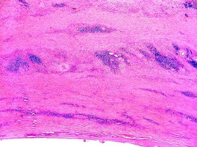
3. A 42-year-old man presents with recurrent low back pain on both sides. He has a history of heavy cigarette smoking and familial hypercholesterolemia, for which he has been receiving appropriate pharmacologic treatment. His erythrocyte sedimentation rate is very elevated. A CT scan of the abdomen reveals a fusiform dilatation of the infrarenal aortic segment, with prominent nodular thickening of its wall. Nodular thickening of the aortic wall appears to extend into the periaortic soft tissues. All major aortic branches are normal. This photomicrograph demonstrates the typical histologic features of this type of aortic aneurysm. Which of the following is the most likely condition associated with this aortic lesion?
QUESTION 30.3
A. Giant cell arteritis
B. IgG4-related systemic disease
C. Kawasaki disease
D. Polyarteritis nodosa
E. Takayasu disease
4. Which of the following forms of vasculitis affects elastic arteries?
A. Churg-Strauss angiitis
B. Henoch-Schönlein purpura
C. Kawasaki disease
D. Polyarteritis nodosa
E. Takayasu disease
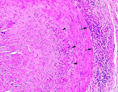
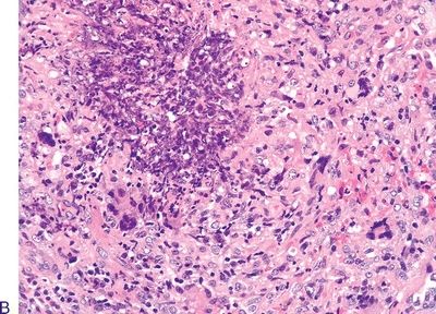
5. A 70-year-old patient presents with temporal headache of acute onset, fever of unknown origin, and a high erythrocyte sedimentation rate. A temporal artery biopsy reveals the changes demonstrated by this photomicrograph. Elastic stain shows near-total destruction of the internal elastic lamina. Which of the following is the most likely diagnosis?
QUESTION 30.5
A. Calcinosis
B. Giant cell arteritis
C. Polyarteritis nodosa
D. Thromboarteritis obliterans (Buerger disease)
E. Wegener granulomatosis
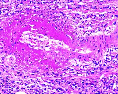
6. A renal biopsy reveals the histopathologic changes shown in this photomicrograph, affecting medium-sized arteries. Vascular lesions appear at different stages of evolution. Laboratory tests indicate a recent hepatitis B infection; antineutrophil cytoplasmic antibodies (ANCAs) are negative. These features are most consistent with:
QUESTION 30.6
A. Churg-Strauss syndrome
B. Giant cell arteritis
C. Leukocytoclastic cutaneous arteritis
D. Polyarteritis nodosa
E. Wegener granulomatosis
7. A 3-year-old boy is brought to the family physician because of a 7-day history of fever unresponsive to over-the-counter medications, bilateral conjunctivitis, strawberry tongue, palmar and plantar erythema with skin peeling, and swollen cervical lymph nodes. This condition is most likely due to:
A. Churg-Strauss syndrome
B. Kawasaki disease
C. Leukocytoclastic cutaneous arteritis
D. Polyarteritis nodosa
E. Wegener granulomatosis
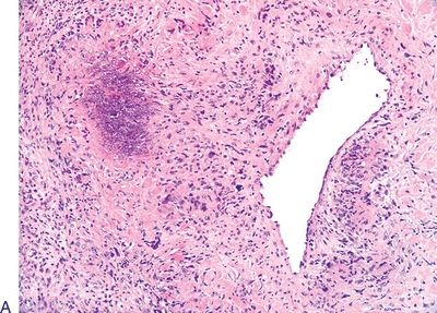
8. A lung biopsy reveals the lesions shown in these pictures. The patient has similar lesions in the sinonasal mucosa and kidneys. Laboratory tests are positive for c-ANCA. Which of the following is the most likely diagnosis?
QUESTION 30.8
A. Churg-Strauss syndrome
Stay updated, free articles. Join our Telegram channel

Full access? Get Clinical Tree


