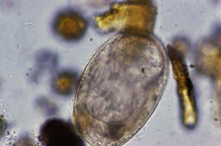
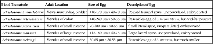
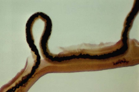
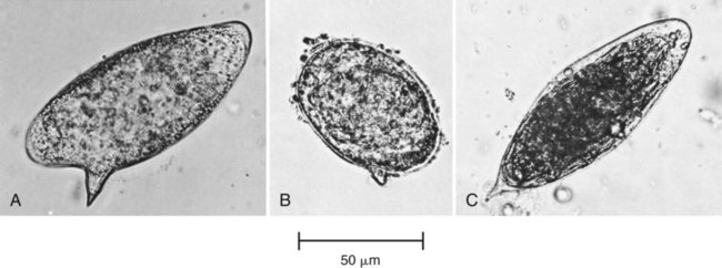
1. List the clinically significant blood trematodes. 2. Describe the general life cycle of the blood trematodes and how human infection occurs. 3. Explain the diagnostic methods used to identify blood trematodes. 4. Differentiate the eggs of the five species of schistosomes. 5. Describe the pathogenesis of the blood trematodes. 6. List the drugs of choice for treatment of blood trematode infections. 7. Describe where blood trematodes are found and how infection may be prevented. Unlike the other trematodes, adult schistosomes are not flattened, but are rather long, thin, and rounded in shape. There is an oral sucker surrounding the mouth and a ventral sucker located just slightly below the oral sucker. The adult male averages 1.5 cm in length and is wider than the female, having a ventral fold that wraps around the female when they mate (Figure 58-1). The adult female averages 2 cm in length and is very thin. The eggs of each species are distinct, and can be distinguished by size, spine morphology, and sometimes specimen type (Figure 58-2). The size range for eggs of S. haematobium is 110 to 170 µm long by 40 to 70 µm wide, and they have a sharply pointed terminal spine. They are fully embryonated without an operculum. The size range for the eggs of S. japonicum is 70 to 100 µm long by 50 to 65 µm wide, and they have a small lateral spine that is sometimes difficult to detect (Figure 58-3). S. mekongi eggs are smaller than those of S. japonicum, ranging in size from 50 to 65 µm long by 30 to 55 µm wide. They are fully embryonated without an operculum and have a small lateral spine. The size range for eggs of S. mansoni is 115 to 180 µm long by 40 to 75 µm wide, and they have a large lateral spine. S. mansoni eggs are unoperculate, immature when released, and take up to 8 to 10 days to develop a miracidium. S. intercalatum eggs are fully embryonated without an operculum, have a terminal spine, and range in size from 140 to 240 µm long by 50 to 85 µm wide. S. intercalatum eggs resemble those of S. haematobium and can be differentiated by Ziehl-Neelsen acid-fast positivity. In addition, S. intercalatum eggs are only found in feces, not in urine specimens. Table 58-1 provides a comparison of the schistosome eggs. TABLE 58-1 Diagnostic Characteristics of the Blood Trematodes
Blood Trematodes
General Characteristics
Blood Trematode
Adult Location
Size of Egg
Description of Egg
Schistosoma haematobium
Veins surrounding bladder
110-170 µm × 40-70 µm
Pointed terminal spine, unoperculated, embryonated
Schistosoma intercalatum
Venules of colon
140-240 µm × 50-85 µm
Resembles egg of S. haematobium, but acid-fast positive
Schistosoma japonicum
Venules of small intestine
70-100 µm × 50-65 µm
Small lateral spine, unoperculated, embryonated
Schistosoma mansoni
Venules of large intestine
115-180 µm × 40-75 µm
Large lateral spine, unoperculated, embryonated
Schistosoma mekongi
Venules of small intestine
50-65 µm × 30-55 µm
Resembles egg of S. mansoni, but much smaller
![]()
Stay updated, free articles. Join our Telegram channel

Full access? Get Clinical Tree


Basicmedical Key
Fastest Basicmedical Insight Engine