


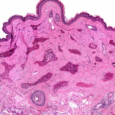
Low magnification of a superficial arteriovenous hemangioma shows a tumor that is mostly composed of thick-walled vessels.
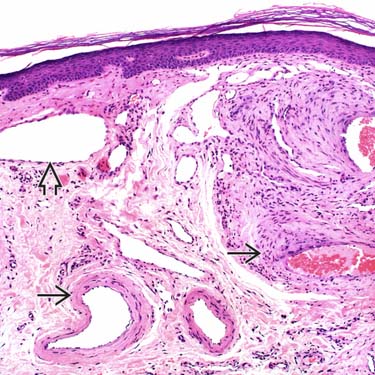
Arteriovenous hemangioma is composed of a circumscribed proliferation of thick-walled muscular vessels that resemble arteries
 and thin-walled ectatic veins
  .
.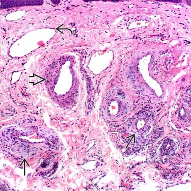
Arteriovenous hemangioma is classically composed of a combination of thick-
 and thin-walled vessels
  . The thick-walled vessels often have degenerative changes resulting in myxoid stroma
  surrounding the vessel.
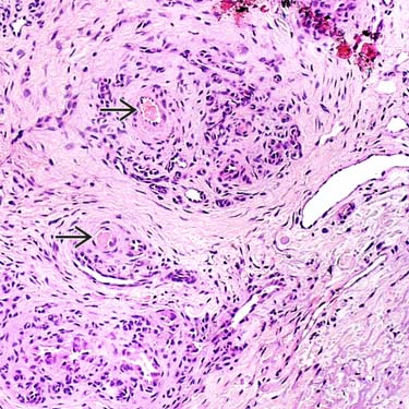
Fibrin thrombi are sometimes present in the lumina of some of the thick-walled vessels, as shown here
 .
.CLINICAL ISSUES
Epidemiology


Site
• Usually involves face


Stay updated, free articles. Join our Telegram channel
Full access? Get Clinical Tree
