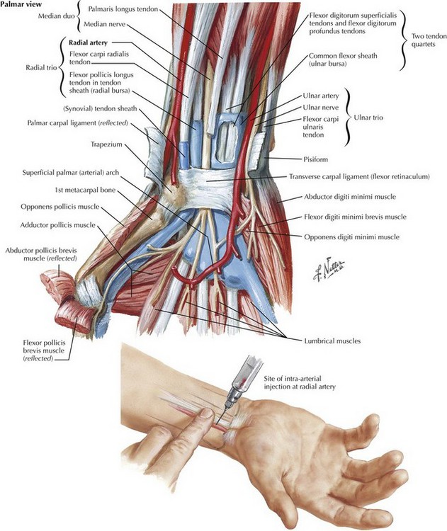
Arterial Line Anatomy

Chapter 41


Arterial Line Anatomy


Radial Artery


The radial artery is chosen most frequently for arterial cannulation because it is readily accessible, relatively easy to cannulate, easily tested for collateral circulation, and associated with few long-term complications (Fig. 41-1). The radial artery is palpated between the distal radius and the flexor carpi radialis tendon. In this area the radial artery is covered by skin, subcutaneous tissue, and fascia only.


Ultrasound can be used to locate the radial artery when it is not palpable. Either a 20-gauge angiocatheter or an arterial line kit may be used, depending on the preference of the practitioner. The catheter is inserted at a 30- to 45-degree angle to the skin until there is pulsatile flow from the artery, at which point the catheter is advanced.


The Allen test is often used to test for collateral circulation using the ulnar artery, although its utility is questionable. To perform this test, the radial and ulnar arteries are manually occluded, preferably after a fist is made by the patient. The ulnar artery is then released, and if collateral circulation is sufficient, perfusion should return to the hand in several seconds.


On occasion, the radial artery is not available for cannulation. In these situations the practitioner must be aware of alternative sites for arterial cannulation, such as the axillary, brachial, femoral, and dorsalis pedis arteries. The technique for cannulation of these sites is similar to that of the radial artery, except that a longer catheter (12 cm) should be used for the axillary, brachial, and femoral sites. Ultrasound may also be helpful in locating these arteries.

Stay updated, free articles. Join our Telegram channel

Aug 12, 2016 | Posted by in ANATOMY | Comments Off on Arterial Line Anatomy

Full access? Get Clinical Tree

Get Clinical Tree app for offline access