Tumors in anogenital area may truly represent adenoma of anogenital mammary-like glands
Tumors in eyelid should be distinguished from endocrine mucin-producing sweat gland carcinoma
• F > M (2:1)
• Adult patients
• Nodule measuring < 2 cm
Sometimes pedunculated
Microscopic
• Circumscribed intradermal nodule
Subcutis may be involved
• Sometimes arises in association with nevus sebaceous and syringocystadenoma papilliferum
• Tubules of different size and shape
• Some cystically dilated
• Papillary projections devoid of fibrovascular cores
True papillae may be seen in cases associated with syringocystadenoma papilliferum
• Outer layer composed of myoepithelial cells
• Inner layer composed of apocrine cells
• Focal follicular or sebaceous differentiation may be rarely seen
Ancillary Tests
• Luminal cells are positive for EMA and CEA
• Myoepithelial cells are positive for S100 and SMA
Top Differential Diagnoses
• Syringocystadenoma papilliferum
• Papillary eccrine adenoma
• Apocrine hamartoma
• Apocrine carcinoma
• Endocrine mucin-producing sweat gland carcinoma
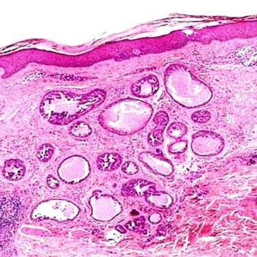
Intradermal Tumor Without Epidermal Connection Apocrine tubular adenoma is usually a well-circumscribed intradermal nodule composed of variably sized and shaped tubules. Some tubular structures are cystically dilated.
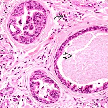
Cystically Dilated Tubules The tubules may contain eosinophilic secretion and debris . The tubular structures are separated by a connective tissue stroma with few blood vessels and scant chronic inflammation.
Tubules Are Lined by Apocrine Cells The tubules are lined by at least 2 layers of cells. The outer layer is composed of small myoepithelial cells , and the luminal layer is composed of columnar apocrine cells with apical snouts.
Apical Snouts in Tubular Apocrine Adenoma Papillary projections are common and usually devoid of fibrovascular cores. Note the decapitation secretions on the surface of the apocrine cells .


 . The tubular structures are separated by a connective tissue stroma with few blood vessels
  and scant chronic inflammation.
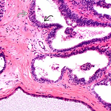
 , and the luminal layer is composed of columnar apocrine cells
  with apical snouts.
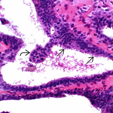
 are common and usually devoid of fibrovascular cores. Note the decapitation secretions on the surface of the apocrine cells
  .
.


