Angiosarcoma
Cyril Fisher, MD, DSc, FRCPath
Key Facts
Terminology
Malignant mesenchymal neoplasm of cells recapitulating variable morphologic and functional features of endothelial cells
Clinical Issues
Deep soft tissues
Lower extremities, followed by upper extremities
Trunk > head and neck
Significant proportion intraabdominal and retroperitoneal
Rare (more frequent in superficial locations)
< 1% of all sarcomas
Any age, but most common in older adults
Poor prognosis irrespective of grade of malignancy
5-year survival 20-30% at best
Aggressive surgical resection with wide tumor-free margins
Microscopic Pathology
Irregular, anastomosing vascular spaces
Variably pleomorphic endothelial tumor cells
Endothelial multilayering and papillary formation
Solid areas common
No complete rim of actin positive myopericytes
Often intracytoplasmic lumina
Prominent nuclear atypia
Numerous mitoses
Expression of endothelial markers
Epithelioid angiosarcomas occur relatively frequently in deep soft tissues
Solid sheets of large epithelioid cells in epithelioid angiosarcoma
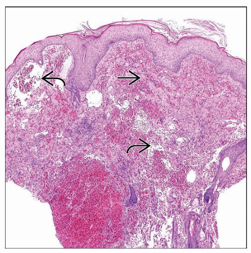 Cutaneous angiosarcoma of the scalp in an elderly man. The dermis contains dilated vascular channels  lined by plump endothelial cells. There are also spindle cell areas lined by plump endothelial cells. There are also spindle cell areas  and focal hemorrhage. and focal hemorrhage. |
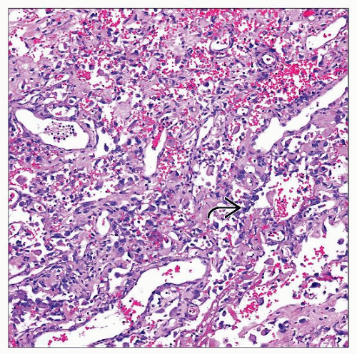 Higher magnification shows well-differentiated angiosarcoma comprising irregularly shaped anastomosing vascular structures with open lumina lined by enlarged, atypical endothelial cells  . . |
TERMINOLOGY
Synonyms
Hemangiosarcoma
Malignant hemangioblastoma
Malignant hemangioendothelioma
Definitions
Malignant mesenchymal neoplasm of cells recapitulating variable morphologic and functional features of endothelial cells
ETIOLOGY/PATHOGENESIS
Developmental Anomaly
Develops rarely in association with genetic syndromes
Klippel-Trenaunay syndrome
Maffucci syndrome
In longstanding congenital lymphedema
Environmental Exposure
Rarely develops adjacent to foreign material or synthetic vascular grafts
In limbs with longstanding lymphedema
After mastectomy: Stewart-Treves syndrome
Post-therapeutic irradiation
CLINICAL ISSUES
Epidemiology
Incidence
Rare; < 1% of all sarcomas
More frequent in superficial locations
1/4 of angiosarcomas arise in deep soft tissues
Age
Occurs at any age, but most common in older adults
Rare subset in childhood
Gender
M > F
Site
Skin of head and neck
Skin of limbs in longstanding lymphedema
Skin of breast following therapeutic irradiation for carcinoma
Deep soft tissue
Lower extremities > upper extremities
Trunk > head/neck region
Significant proportion arises in abdomen and retroperitoneum
Rarely multifocal
Presentation
Slow growing
Deep mass
Usually large mass
Hematologic abnormalities
Thrombocytopenia may be present
Arteriovenous shunting may be present
Rarely arises in nonlipogenic component of dedifferentiated liposarcomas
Rarely arises in benign or malignant nerve sheath tumors
Very rarely arises in preexisting benign hemangioma
Skin lesions
Plaque or nodule with purple discoloration and bruising
Treatment
Surgical approaches
Aggressive surgical resection with wide tumor-free margins
Adjuvant therapy
Response to chemotherapy
Inhibition of angiogenesis
MACROSCOPIC FEATURES
General Features
Infiltrating neoplasm
Areas of hemorrhage
MICROSCOPIC PATHOLOGY
Histologic Features
Angiosarcoma
Usually no relationship to preexisting vessels
Irregular infiltrating and anastomosing vascular spaces
Variably pleomorphic endothelial tumor cells
Nuclear atypia and prominent nucleoli
Endothelial multilayering and papillary formation
Solid areas common
Neoplastic vascular structures encircled by reticulin fibers
No complete rim of SMA positive (myo)pericytes
Often intracytoplasmic lumina that may contain erythrocytes
Mitoses are usually numerous
Areas of hemorrhage and necrosis may be present
Clear distinction between lymphatic and vascular differentiation remains problematic
Epithelioid angiosarcoma
In cutaneous or, more commonly, in deep soft tissues
Often rapid growth
Very aggressive clinical course
Solid sheets of large epithelioid cells
Tumor cells with abundant eosinophilic cytoplasm and large vesicular nuclei
Prominent cytologic atypia and numerous mitoses
Often areas of tumor necrosis
Rare predominantly spindle cell morphology
Stay updated, free articles. Join our Telegram channel

Full access? Get Clinical Tree


