• Admixture of mature adipocytes and capillary-sized blood vessels
Hallmark: Fibrin thrombi within vessels
• Vascularity often more prominent in periphery
• No nuclear atypia in adipocytic or endothelial components
• Morphologic variant: Cellular angiolipoma
Top Differential Diagnoses
• Lipoma
• Intramuscular hemangioma
• Kaposi sarcoma
• Spindle cell hemangioma
• Angiomyolipoma
Angiolipoma at Low Magnification Angiolipoma is composed of both vascular and mature adipocytic components admixed together in varying proportions. They are often small, circumscribed lesions, as seen here.
Prominent Adipose Tissue Component This angiolipoma is predominantly composed of adipose tissue. The focal vascular component is present as clusters of small vessels near the periphery and around the thin, fibrous septa .
Peripheral Vascular Component The focal vascular component of angiolipoma is present as clusters of small vessels near the periphery of the lesion . The remainder of the tumor is composed of mature adipocytes .
Characteristic Fibrin Thrombi Bright pink fibrin thrombi are present in small clustered capillary vessels. This is the hallmark feature of angiolipoma.
TERMINOLOGY
Definitions
• Benign tumor composed of mature adipocytes and clustered small blood vessels with intraluminal fibrin thrombi
ETIOLOGY/PATHOGENESIS
Unknown
• Majority are sporadic
• Rare familial predilection (5%)
CLINICAL ISSUES
Epidemiology
• Age
Most common in young adults
Rare in children or in adults > 50 years
• Sex
Male predominance
Site
• Forearm, trunk, and upper arm most common sites
• Rare on scalp or face
• May occur in subcutis of breast (causing clinical concern for neoplasm of breast parenchyma)
Presentation
• Painful subcutaneous nodule, often multiple (in 2/3 of cases)
No correlation between pain and degree of tumor vascularity
Treatment
• Simple surgical excision
Prognosis
• Excellent: Benign
Very low risk of local recurrence
No known risk of malignant transformation
MACROSCOPIC
Gross Features
• Well circumscribed
• Yellow-red nodules
• Typically < 2 cm in size
MICROSCOPIC
Histologic Features
• 2 components present in varying proportions
Mature adipocytes
Clustered capillary-sized vessels with fibrinoid thrombi
– Vascularity often more prominent in periphery
• No nuclear atypia in adipocytic or endothelial components
• Fibrosis may be present around vessels and between adipocytes in late stage of lesion
• Mast cells may be conspicuous in some cases
Morphologic Variant
• Cellular angiolipoma
Vascular component predominates
Only gold members can continue reading. Log In or Register to continue


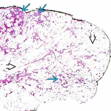
 and mature adipocytic
  components admixed together in varying proportions. They are often small, circumscribed lesions, as seen here.
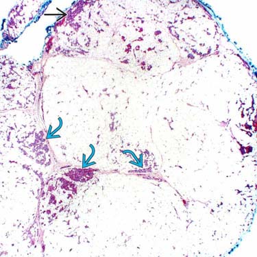
 and around the thin, fibrous septa
  .
.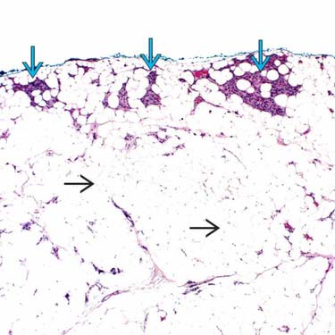
 . The remainder of the tumor is composed of mature adipocytes
  .
.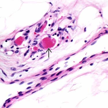
 are present in small clustered capillary vessels. This is the hallmark feature of angiolipoma.


