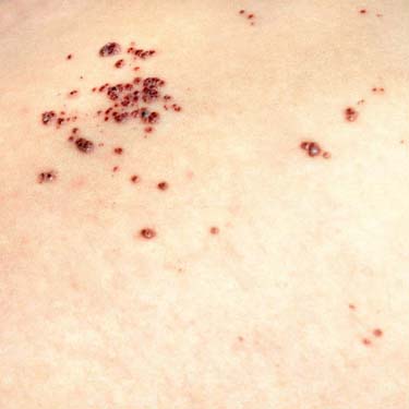
Clinical Image of Angiokeratomas Clinical image demonstrates numerous grouped angiokeratomas in a patient with Anderson-Fabry disease.
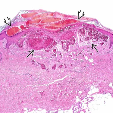
Angiokeratoma at Low Magnification Angiokeratomas are characterized by ectatic vessels in the superficial dermis that appear to herniate into the epidermis. There is often overlying hemorrhage , epidermal acanthosis, and frequently compact hyperkeratosis.
Angiokeratoma With Epidermal Acanthosis and Hyperkeratosis The epidermis overlying angiokeratomas typically demonstrates acanthosis and compact hyperkeratosis . Numerous, dilated, superficial dermal blood vessels are also seen in this field.
Angiokeratoma at High Magnification The vessels of angiokeratoma herniate up into the epidermis . The vessels frequently undergo thrombosis, and in this case demonstrates an early thrombus characterized by fibrin admixed with erythrocytes.


 in the superficial dermis that appear to herniate into the epidermis. There is often overlying hemorrhage
  , epidermal acanthosis, and frequently compact hyperkeratosis.
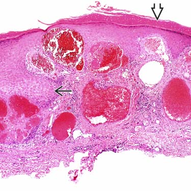
 and compact hyperkeratosis
  . Numerous, dilated, superficial dermal blood vessels are also seen in this field.
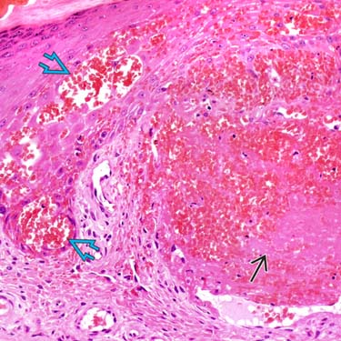
 . The vessels frequently undergo thrombosis, and in this case demonstrates an early thrombus characterized by fibrin
  admixed with erythrocytes.


