Solitary, dome-shaped, flesh-colored papules on nose or central face

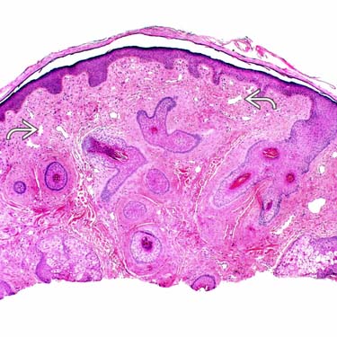
Fibrous papule (FP) is characterized by a dome-shaped lesion with ectatic thin-walled blood vessels
 and a dense collagenous stroma.
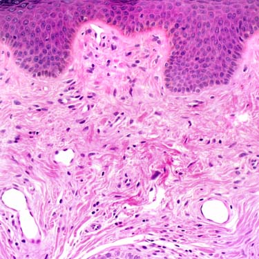
This higher power image of an FP demonstrates the relatively hypocellular proliferation of bland spindled to stellate fibroblasts, small ectatic vessels, and dense collagenous stroma.

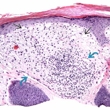
This example of clear cell FP is composed of epithelioid cells with abundant clear-staining cytoplasm
 . The lesion also has an associated mildly collagenous stroma and a few small, dilated vessels
  .
.
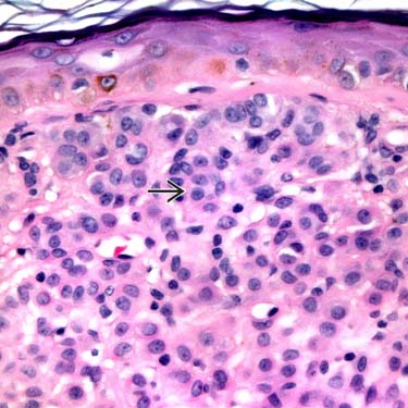
Epithelioid FP is more cellular and shows a proliferation of round cells with eosinophilic cytoplasm
 .
.TERMINOLOGY
Synonyms
Definitions
• Angiofibroma encompasses group of benign mesenchymal tumors characterized by spindled to stellate fibroblasts, dense collagenous stroma, and ectatic blood vessels


Stay updated, free articles. Join our Telegram channel

Full access? Get Clinical Tree


