Anatomy of the Respiratory System
LANGUAGE OF SCIENCE
 Before reading the chapter, say each of these terms out loud. This will help you avoid stumbling over them as you read.
[alve- hollow, -ol- little, -ar relating to]
[alve- hollow, -olus little] pl., alveoli
[bronch- windpipe, -al relating to]
[bronch- windpipe, -ol- little]
[bronch- windpipe, -pulmon- lung, -ary relating to]
[concha sea shell] pl., conchae
[epi- upon, -glottis tongue] pl., epiglottides or epiglottises
[glottis tongue] pl., glottides or glottises
[laryng- voicebox (larynx), -pharynx throat] pl., laryngopharynges or laryngopharynxes
[larynx voicebox] pl., larynges or larynxes
[ling- tongue, -al relating to]
[re- again, -spir- breathe, -tory relating to, tractus trail]
[nas- nose, -al relating to, mucus slime] pl., mucosae
[naso- nose, -pharynx throat] pl., nasopharynges or nasopharynxes
[obliqu- slanted, fissur- cleft]
(ohl-FAK-tor-ee ep-ih-THEE-lee-um)
[olfact- smell, -ory relating to, epi– upon, –theli- nipple, -um thing] pl., epithelia
[oro- mouth, -pharynx throat] pl., oropharynges or oropharynxes
[palat- palate, -ine relating to]
[para- beside, -nas- nose, -al relating to, sinus hollow]
[parie- wall, -al relating to, pleura rib] pl., pleurae
[pharyng- throat, -al relating to]
[pharynx throat] pl., pharynges or pharynxes
[prim- first, -ary relating to, bronchus windpipe] pl., bronchi
[re- again, -spir- breathe, -tory relating to, membran- thin skin]
(RES-pih-rah-tor-ee myoo-KOH-sah)
[re- again, -spir- breathe, -tory relating to, mucus slime]
[re- again, -spir- breathe, -tory relating to]
[second- second, bronchus windpipe] pl., bronchi
[combination of surf(ace) act(ive) a(ge)nt]
[trachea rough duct] pl., tracheae or tracheas
[turbin- top (spinning toy), -ate of or like]
[re- again, -spir- breathe, -tory relating to, tract- trail]
[vestibul- entrance hall, voca- voice, -al relating to]
[vibrissa nostril hair] pl., vibrissae
[viscer- internal organ, -al relating to, pleura rib] pl., pleurae
[voca- voice, -al relating to]
LANGUAGE OF MEDICINE
[acut- sharp, bronch- windpipe, -itis inflammation]
chronic obstructive pulmonary disease (COPD)
(KRON-ik ob-STRUK-tiv PUL-moh-nair-ee)
[chron- time, -ic relating to, pulmon- lung, -ary relating to]
[devia- turn aside, -ate process, septum partition] pl., septa
(en-doh-TRAY-kee-al in-too-BAY-shun)
[endo- within, -trache- rough duct, -al relating to, in- within, -tub- tube, -ation process]
[epi– upon, -glotti- tongue (glottis), -itis inflammation]
[laryng- voice box (larynx), -itis inflammation]
[named for American Legion convention, location of first diagnosed case]
[cancer crab or malignant tumor]
[oti- ear, -itis inflammation, media middle]
[pharyng- throat (pharynx), -itis inflammation]
[photo- light, -dynam- force, -ic relating to]
[pneumon- lung, –ec- out, -tom- cut, -y action]
[rhin- nose, -itis inflammation]
[sinus- hollow, -itis inflammation]
[tonsil- tonsil, –ec- out, -tom- cut, -y action]
[tonsil- tonsil, litis inflammation]
[trache- rough duct, -os- mouth or opening, -tom- cut, -y action]
[tuber- swelling, -cul- little, -osis condition]
upper respiratory infection (URI)
[re- again, -spir- breathe, -tory relating to, infec- stain, -tion process]
STRUCTURAL PLAN OF THE RESPIRATORY SYSTEM
For purposes of study, the respiratory system may be divided into upper and lower tracts, or structural divisions. The organs of the upper respiratory tract are located outside the thorax, or chest cavity, whereas those in the lower tract, or division, are located almost entirely within it (Figure 26-1).
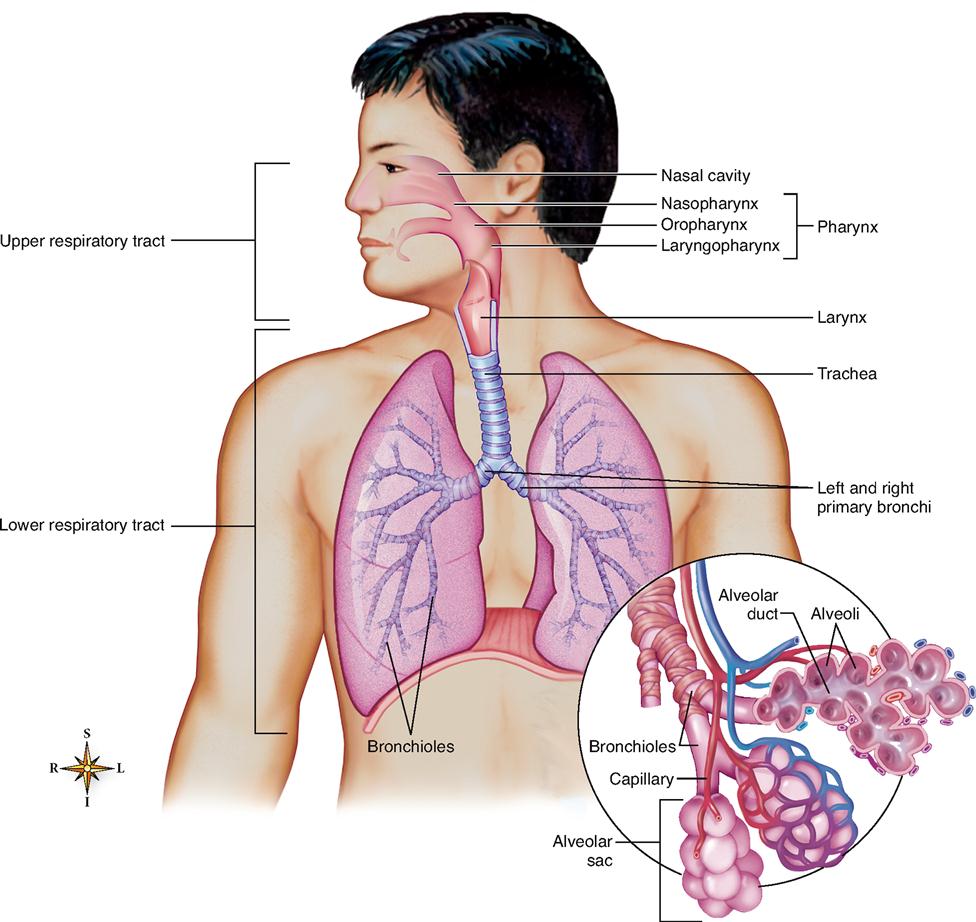
The upper respiratory tract is composed of the nose, nasopharynx, oropharynx, laryngopharynx, and larynx. The lower respiratory tract, or division, consists of the trachea, all segments of the bronchial tree, and the lungs. Functionally, the respiratory system also includes several accessory structures, such as the oral cavity, rib cage, and respiratory muscles including the diaphragm. Together, these structures constitute the lifeline, the air supply line of the body. This chapter describes the functional anatomy of these organs. The physiology of the respiratory system as a whole is discussed in Chapter 27. Cells require a constant supply of oxygen for the vital energy conversion process carried out within each cell’s mitochondria—a process called cellular respiration (Chapter 30). Cellular respiration produces carbon dioxide (CO2) as a waste product, which must be removed before it accumulates to dangerously high levels.
UPPER RESPIRATORY TRACT
Nose
STRUCTURE OF THE NOSE
The nose has external and internal structures. The external portion, that is, the part that protrudes from the face, consists of a bony and cartilaginous framework overlaid by skin containing many sebaceous glands (Box 26-1). The two nasal bones meet in the center of the face just below the forehead, where they are surrounded by the frontal bone to form the root of the nose. The nose is surrounded by the maxilla laterally and inferiorly at its base. The flaring cartilaginous expansion forming and supporting the outer side of each oval nostril opening is called the ala.
Box 26-1
 FYI
FYIDanger Area of the Face
The fact that the skin of the external nose contains many sebaceous glands has great clinical significance. If these glands become infected, it is possible for infectious material to enter and pass from facial veins near the nose to one of the intracranial venous sinuses (see Chapter 21). For this reason, the triangle-shaped zone surrounding the external nose is often known as the “danger area of the face.”
The internal portion of the nose, or nasal cavity, lies over the roof of the mouth where the palatine bones, which form the floor of the nose and the roof of the mouth, separate the nasal cavities from the mouth cavity. Sometimes the palatine bones fail to unite completely and produce a condition known as cleft palate. When this abnormality exists, the mouth is only partially separated from the nasal cavity, and consequently, difficulties arise in swallowing and speaking.
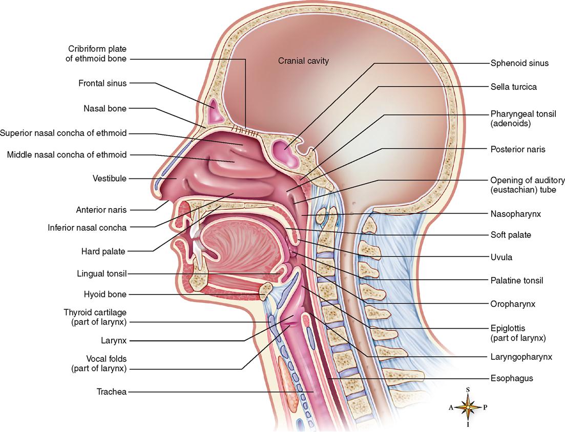
The roof of the nose is separated from the cranial cavity by a portion of the ethmoid bone called the cribriform plate (Figures 26-2 and 26-3). The cribriform plate is perforated by many small openings that permit branches of the olfactory nerve responsible for the special sense of smell to enter the cranial cavity and reach the brain.
Separation of the nasal and cranial cavities by a thin, perforated plate of bone presents real hazards. If the cribriform plate is damaged as a result of trauma to the nose, it is possible for potentially infectious material to pass directly from the nasal cavity into the cranial fossa and infect the brain and its covering membranes.
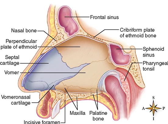
The hollow nasal cavity is separated by a midline partition, the septum (see Figure 26-2), into right and left cavities. Note in Figure 26-2 that the nasal septum is made up of four main structures: the perpendicular plate of the ethmoid bone above, the vomer bone, and the septal nasal and vomeronasal cartilages below. In the adult the nasal septum is frequently deviated to one side or the other, interfering with respiration and with drainage of the nose and sinuses. The nasal septum has a rich blood supply. Nosebleeds, or epistaxis, often occur as a result of septal contusions caused by a direct blow to the nose. Epistaxis may also result from weak blood vessels combined with high blood pressure. Though seemingly dramatic, nosebleeds are seldom a serious health problem.
Each nasal cavity is divided into three passageways named the superior, middle, and inferior meatuses. These incomplete tubes are formed by the projection of the conchae, or turbinates, curving from the lateral walls of the internal portion of the nose (see Figure 26-3). The superior and middle conchae are processes of the ethmoid bone, whereas the inferior conchae are separate bones.
The external openings into the nasal cavities, commonly referred to as nostrils, have the technical name of anterior nares (singular, naris). They are sometimes called external nares. The anterior nares open into an area covered by skin that is reflected from the wings (ala) of the nose. This area, called the vestibule, is located just inside the nasal cavity below the inferior meatus. The vestibule is lined with skin. Coarse hairs called vibrissae, sebaceous glands, and numerous sweat glands are found in the skin of the vestibule. Once air has passed over the skin of the vestibule, it enters the respiratory portion of each nasal passage. This area extends from the inferior meatus to the small funnel-shaped orifices of the posterior (internal) nares. The posterior nares are openings that allow air to pass from the nasal cavity into the next major segment of the upper respiratory tract—the pharynx.
If one were to “trace” the movement of air through the nose into the pharynx, it would be found that air passes through several structures on the way. The sequence is as follows:
Nasal Mucosa
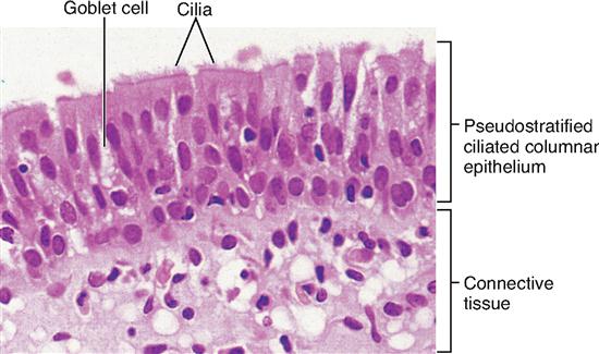
Once air has passed over the skin of the vestibule and enters the respiratory portion of the nasal passage, it passes over the respiratory mucosa. This mucous membrane has a pseudostratified ciliated columnar epithelium rich in goblet cells (Figure 26-4). The respiratory mucosa possesses a rich blood supply, especially over the inferior turbinate, and is bright pink or red. Near the roof of the nasal cavity and over the superior turbinate and opposing portion of the septum, the mucosa turns pale and has a yellowish tint. In this area it is referred to as the olfactory epithelium. This membrane contains many olfactory nerve cells and has a rich lymphatic plexus. Ciliated mucous membrane lines the rest of the respiratory tract down as far as the smaller bronchioles.
Besides the olfactory sensory neurons imbedded in the nasal mucosa, a structure called the vomeronasal organ (VNO) is located in the mucosa of the inferior nasal septum. The VNO is thought to be involved in sensing pheromones, which are sex signaling chemicals. Want to know more? Check out Pheromones and the Vomeronasal Organ and Nerve Zero online at A&P Connect.

Paranasal Sinuses
The four pairs of paranasal sinuses are air-containing spaces that lighten the weight of the skull and open, or drain, into the nasal cavity. They take their names from the skull bones in which they are located (see Chapter 9). These paranasal sinuses are the frontal, maxillary, ethmoid, and sphenoid sinuses (Figure 26-5). Like the nasal cavity, each paranasal sinus is lined by respiratory mucosa. The mucous secretions produced in the sinuses are continually being swept into the nose by the ciliated surface of the respiratory membrane.
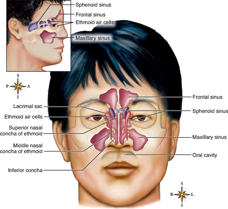
The right and left frontal sinuses are located just above the corresponding orbit, whereas the maxillary, the largest of the sinuses, extends into the maxilla on either side of the nose. The sphenoid sinuses lie in the body of the sphenoid bone on either side of the midline in close proximity to the optic nerves and pituitary gland.
Note in Figure 26-5 that the ethmoid sinuses are not single large cavities but a collection of small air cells divided into anterior, middle, and posterior groups that open independently into the upper part of the nasal cavity (Box 26-2). The paranasal sinuses drain as follows:

 Into the superior meatus—posterior ethmoidal sinuses
 Into the space above the superior conchae (sphenoethmoidal recess)—sphenoid sinuses
 FYI
FYIFUNCTIONS OF THE NOSE
The nose serves as a passageway for air going to and from the lungs. However, if the nasal passages are obstructed, it is possible for air to bypass the nose and enter the respiratory tract directly through the mouth.
Air that enters the system through the nasal cavity is filtered of impurities, warmed, moistened, and chemically examined (by olfaction) to detect substances that might prove irritating to the delicate lining of the respiratory tract. The vibrissae, or nasal hairs, in the vestibule serve as an initial “filter” that screens particulate matter from air that is entering the system. The conchae, or turbinates, then serve as baffles to slow and stir the air—as well as provide a large mucus-covered surface area over which air must pass before reaching the pharynx. The respiratory membrane produces copious quantities of mucus and possesses a rich blood supply, especially over the inferior conchae, which permits rapid warming and moistening of the dry inspired air. Mucous secretions provide the final “trap” where some of the remaining particulate matter from air is removed as it travels through the nasal passages. Fluid from the lacrimal glands (see Figure 17-19 on p. 525) and additional mucus produced in the paranasal sinuses also help trap particulate matter and moisten air passing through the nose.
In addition, the hollow sinuses act to lighten the bones of the skull and serve as resonating chambers for speech. Swirling of air by the middle and superior conchae over the olfactory epithelium makes the special sense of olfaction possible.
Pharynx
STRUCTURE OF THE PHARYNX
Another name for the pharynx is the throat. It is a tubelike structure about 12.5 cm (5 inches) long that extends from the base of the skull to the esophagus and lies just anterior to the cervical vertebrae. It is made of muscle and is lined with mucous membrane.
The pharynx has three anatomical divisions. The nasopharynx is located behind the nose and extends from the posterior nares to the level of the soft palate. The oropharynx is located behind the mouth from the soft palate above to the level of the hyoid bone below. Finally, the laryngopharynx extends from the hyoid bone to the esophagus. Figure 26-3 shows the divisions of the pharynx.
Seven openings are found in the pharynx (see Figure 26-3):


