• Seen in 50-100% of patients with familial adenomatous polyposis and Gardner syndrome
Most common site of extracolonic polyps in syndromic patients
• Signs and symptoms of biliary or pancreatic obstruction
• Precursor of ampullary adenocarcinoma
124x increased risk in syndromic patients
• Endoscopic findings
Soft polyp or plaque with regular margins
Prominence or mucosal thickening of papilla
Absence of ulceration or spontaneous bleeding
May extend into distal bile duct &/or pancreatic duct
Microscopic
• Tubular adenoma
Simple tubular glands resembling basal portions of normal intestinal crypts
< 25% of villous component
• Tubulovillous adenoma
> 25% of both tubular and villous components
• Villous adenoma
> 75% of villous component
Usually sessile
• Areas of high-grade dysplasia may be present
Top Differential Diagnoses
• Reactive epithelial atypia
• Invasive ampullary adenocarcinoma
Extension of adenomatous epithelium into underlying periampullary glands and ducts may mimic invasion
• Flat intraepithelial neoplasia (dysplasia)
• Noninvasive papillary neoplasm, pancreaticobiliary type
Gross Appearance This surgically resected specimen shows a well-circumscribed mass lesion occupying the ampulla of Vater that obstructs the common bile duct and pancreatic duct. Histologic examination shows tubulovillous adenoma with associated invasive adenocarcinoma.
Tubular Adenoma This polypoid ampullary lesion is a tubular adenoma. Note the extension of adenomatous epithelium into the underlying periampullary glands/ducts .
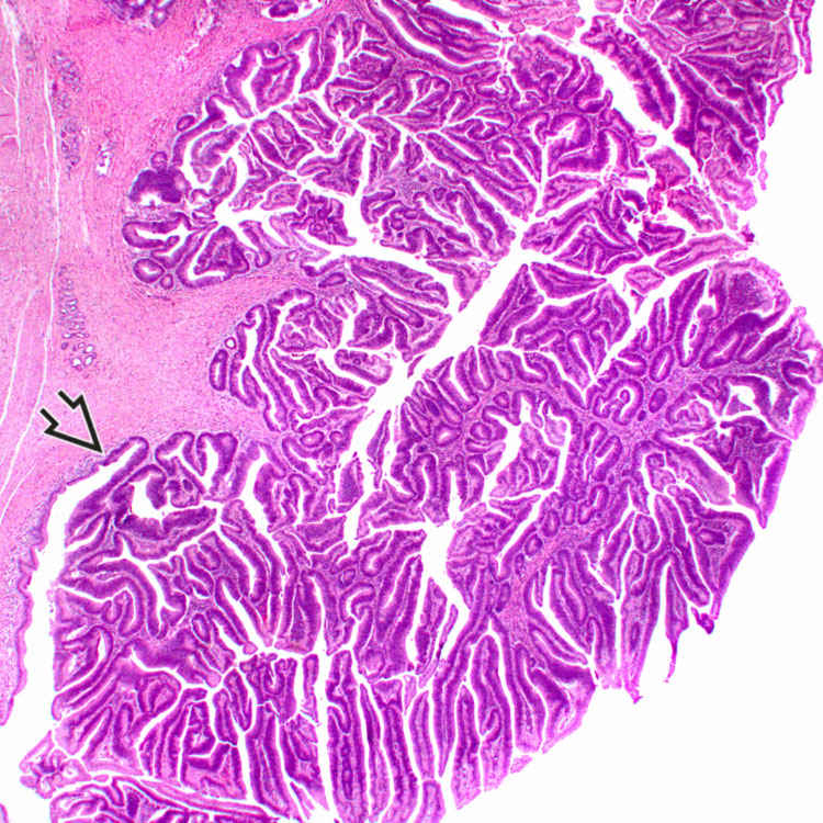
Villous Adenoma This ampullary lesion is a villous adenoma. It extends into the distal common bile duct and shows a continuum of the adenomatous epithelium with the nonneoplastic epithelium lining the bile duct .
Histologic Features This ampullary adenoma shows predominantly basally located nuclei that are elongated, hyperchromatic, and pseudostratified. Numerous Paneth cells are present in this case. Goblet cells and endocrine cells are also present. A few apoptotic bodies are seen.
TERMINOLOGY
Definitions
• Intestinal-type premalignant epithelial neoplastic lesion of ampulla of Vater
CLINICAL ISSUES
Epidemiology
• Incidence
0.04-0.12% of individuals based on autopsy data
Only gold members can continue reading. Log In or Register to continue


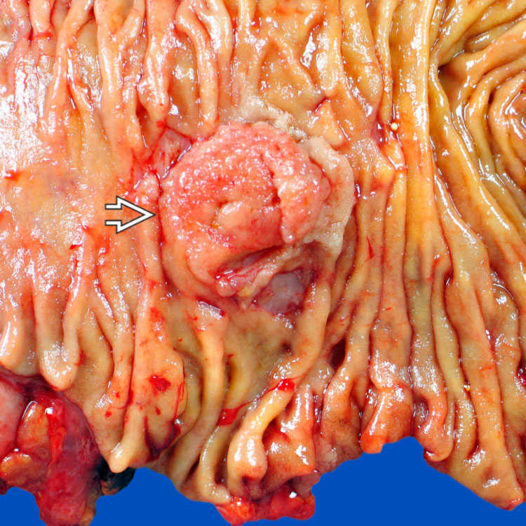
 occupying the ampulla of Vater that obstructs the common bile duct and pancreatic duct. Histologic examination shows tubulovillous adenoma with associated invasive adenocarcinoma.
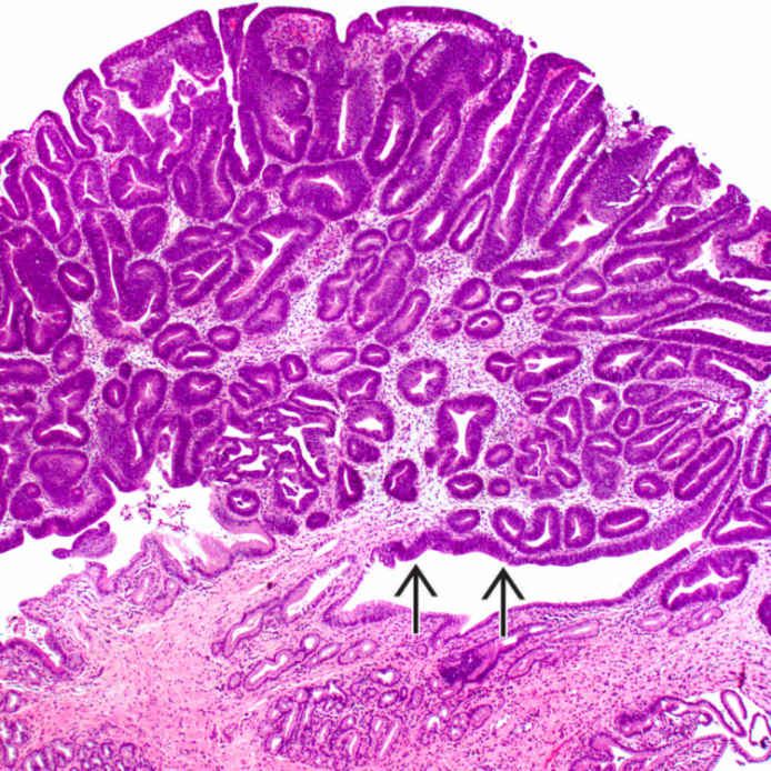
 .
.
 .
.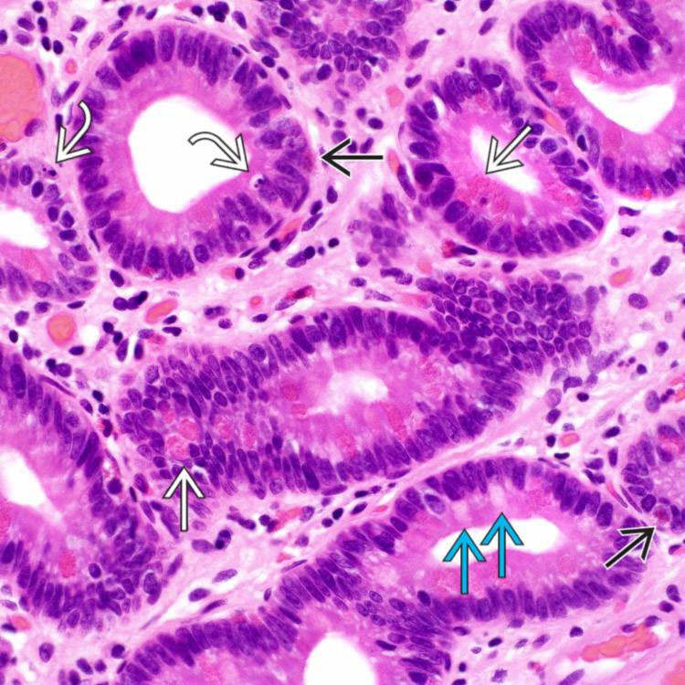
 are present in this case. Goblet cells
  and endocrine cells
  are also present. A few apoptotic bodies
  are seen.


