447 | Migraine and Other Primary Headache Disorders |
The general principles around headache as a cardinal symptom are covered elsewhere (Chap. 21); here we discuss disorders in which headache and associated features occur in the absence of any exogenous cause. The most common are migraine, tension-type headache, and the trigeminal autonomic cephalalgias, notably cluster headache; the complete list is summarized in Table 447-1.
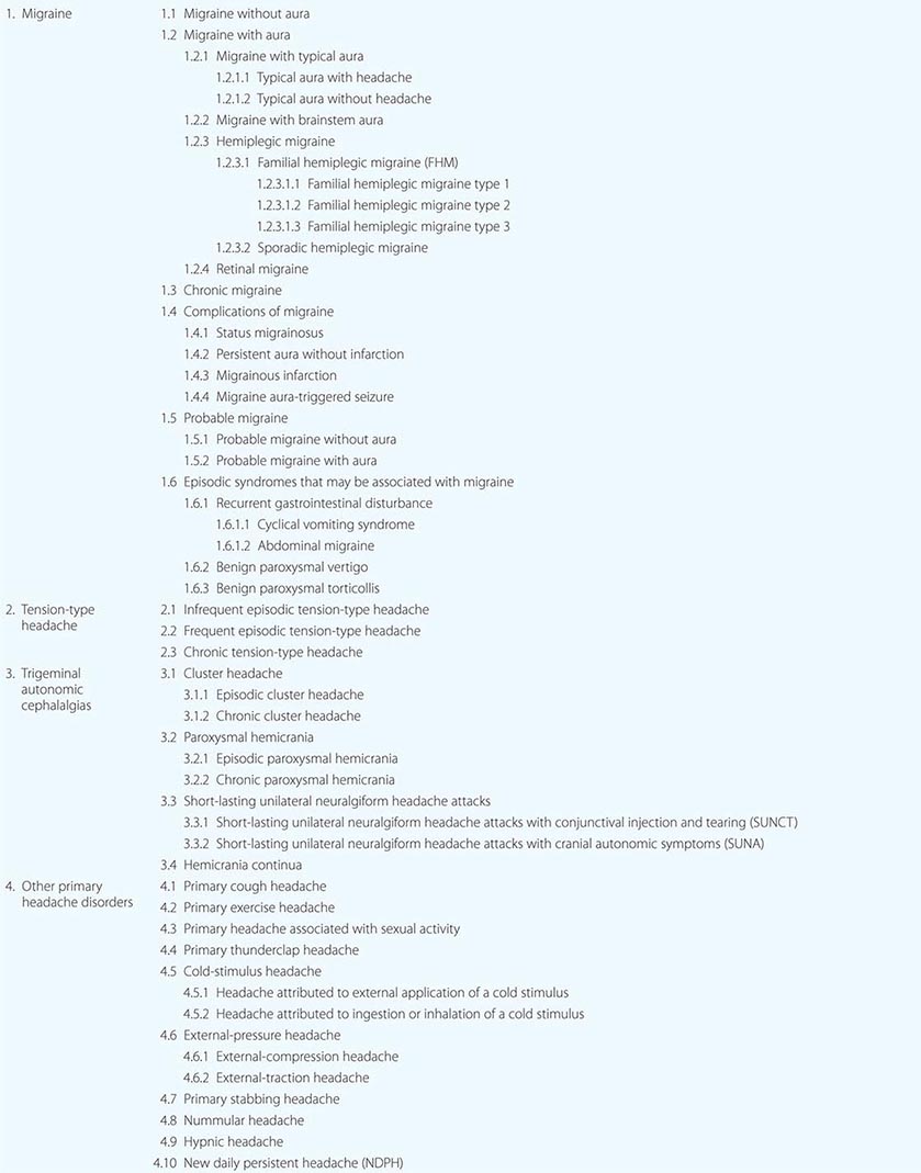
PRIMARY HEADACHE DISORDERS, MODIFIED FROM INTERNATIONAL CLASSIFICATION OF HEADACHE DISORDERS-III-BETA (HEADACHE CLASSIFICATION COMMITTEE OF THE INTERNATIONAL HEADACHE SOCIETY, 2013) |
MIGRAINE
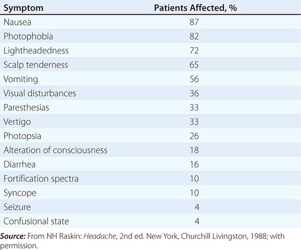
Migraine, the second most common cause of headache, and the most common headache-related, and indeed neurologic, cause of disability in the world, afflicts approximately 15% of women and 6% of men over a 1-year period. It is usually an episodic headache associated with certain features such as sensitivity to light, sound, or movement; nausea and vomiting often accompany the headache. A useful description of migraine is a recurring syndrome of headache associated with other symptoms of neurologic dysfunction in varying admixtures (Table 447-2). Migraine can often be recognized by its activators, referred to as triggers.
SYMPTOMS ACCOMPANYING SEVERE MIGRAINE ATTACKS IN 500 PATIENTS |
The brain of the migraineur is particularly sensitive to environmental and sensory stimuli; migraine-prone patients do not habituate easily to sensory stimuli. This sensitivity is amplified in females during the menstrual cycle. Headache can be initiated or amplified by various triggers, including glare, bright lights, sounds, or other afferent stimulation; hunger; let-down from stress; physical exertion; stormy weather or barometric pressure changes; hormonal fluctuations during menses; lack of or excess sleep; and alcohol or other chemical stimulation, such as with nitrates. Knowledge of a patient’s susceptibility to specific triggers can be useful in management strategies involving lifestyle adjustments.
Pathogenesis The sensory sensitivity that is characteristic of migraine is probably due to dysfunction of monoaminergic sensory control systems located in the brainstem and hypothalamus (Fig. 447-1).
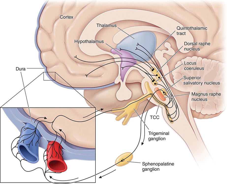
FIGURE 447-1 Brainstem pathways that modulate sensory input. The key pathway for pain in migraine is the trigeminovascular input from the meningeal vessels, which passes through the trigeminal ganglion and synapses on second-order neurons in the trigeminocervical complex (TCC). These neurons in turn project in the quintothalamic tract and, after decussating in the brainstem, synapse on neurons in the thalamus. Important modulation of the trigeminovascular nociceptive input comes from the dorsal raphe nucleus, locus coeruleus, and nucleus raphe magnus.
Activation of cells in the trigeminal nucleus results in the release of vasoactive neuropeptides, particularly calcitonin gene–related peptide (CGRP), at vascular terminations of the trigeminal nerve and within the trigeminal nucleus. CGRP receptor antagonists, gepants, have now been shown to be effective in the acute treatment of migraine, and monoclonal antibodies to CGRP have been shown effective in two early phase clinical trials. Centrally, the second-order trigeminal neurons cross the midline and project to ventrobasal and posterior nuclei of the thalamus for further processing. Additionally, there are projections to the periaqueductal gray and hypothalamus, from which reciprocal descending systems have established antinociceptive effects. Other brainstem regions likely to be involved in descending modulation of trigeminal pain include the nucleus locus coeruleus in the pons and the rostroventromedial medulla.
Pharmacologic and other data point to the involvement of the neurotransmitter 5-hydroxytryptamine (5-HT; also known as serotonin) in migraines. Approximately 60 years ago, methysergide was found to antagonize certain peripheral actions of 5-HT and was introduced as the first drug capable of preventing migraine attacks. The triptans were designed to stimulate selectively subpopulations of 5-HT receptors; at least 14 different 5-HT receptors exist in humans. The triptans are potent agonists of 5-HT1B and 5-HT1D receptors, and some are active at the 5-HT1F receptors; the latter’s exclusive agonists are called ditans. Triptans arrest nerve signaling in the nociceptive pathways of the trigeminovascular system, at least in the trigeminal nucleus caudalis and trigeminal sensory thalamus, in addition to cranial vasoconstriction, while ditans, now shown conclusively to be effective in acute migraine, act only at neural targets. An interesting range of neural targets is now being actively pursed for the acute and preventive management of migraine.
Data also support a role for dopamine in the pathophysiology of migraine. Most migraine symptoms can be induced by dopaminergic stimulation. Moreover, there is dopamine receptor hypersensitivity in migraineurs, as demonstrated by the induction of yawning, nausea, vomiting, hypotension, and other symptoms of a migraine attack by dopaminergic agonists at doses that do not affect nonmigraineurs. Dopamine receptor antagonists are effective therapeutic agents in migraine, especially when given parenterally or concurrently with other antimigraine agents. Moreover, hypothalamic activation, anterior to that seen in cluster headache, has now been shown in the premonitory phase of migraine using functional imaging, and this may hold a key to understanding some part of the role of dopamine in the disorder.
Migraine genes identified by studying families with familial hemiplegic migraine (FHM) reveal involvement of ion channels, suggesting that alterations in membrane excitability can predispose to migraine. Mutations involving the Cav2.1 (P/Q)–type voltage-gated calcium channel CACNA1A gene are now known to cause FHM 1; this mutation is responsible for about 50% of FHMs. Mutations in the Na+-K+ATPase ATP1A2 gene, designated FHM 2, are responsible for about 20% of FHMs. Mutations in the neuronal voltage-gated sodium channel SCN1A cause FHM 3. Functional neuroimaging has suggested that brainstem regions in migraine (Fig. 447-2) and the posterior hypothalamic gray matter region close to the human circadian pacemaker cells of the suprachiasmatic nucleus in cluster headache (Fig. 447-3) are good candidates for specific involvement in primary headache.
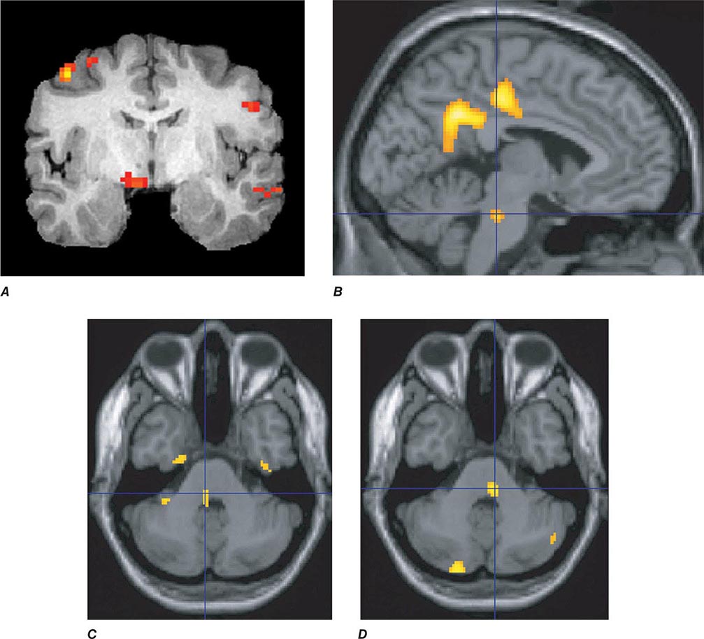
FIGURE 447-2 Positron emission tomography (PET) activation in migraine. Hypothalamic, dorsal midbrain, and dorsolateral pontine activation is seen in triggered attacks in the premonitory phase before pain, whereas in migraine attacks, dorsolateral pontine activation persists, as it does in chronic migraine (not shown). The dorsolateral pontine area, which includes the noradrenergic locus coeruleus, is fundamental to the expression of migraine. Moreover, lateralization of changes in this region of the brainstem correlates with lateralization of the head pain in hemicranial migraine; the scans shown in panels C and D are of patients with acute migraine headache on the right and left side, respectively. (Panel A from FH Maniyar et al: Brain 137:232, 2014; panel B from SK Afridi et al: Arch Neurol 2005;62:1270; Panels C and D from SK Afridi et al: Brain 128:932, 2005.)
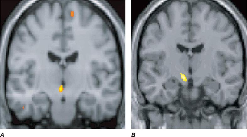
FIGURE 447-3 A. Posterior hypothalamic gray matter activation by positron emission tomography in a patient with acute cluster headache. (From A May et al: Lancet 352:275, 1998.) B. High-resolution T1-weighted magnetic resonance image obtained using voxel-based morphometry demonstrates increased gray matter activity, lateralized to the side of pain in a patient with cluster headache. (From A May et al: Nat Med 5:836, 1999.)
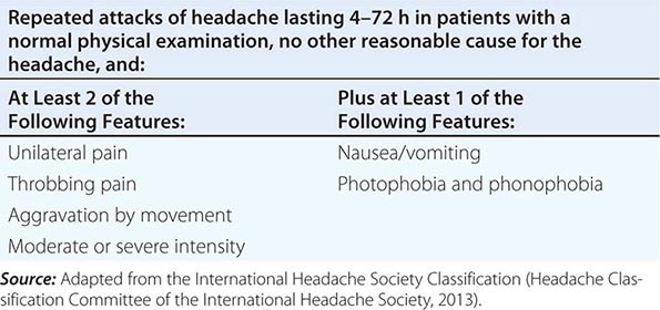
Diagnosis and Clinical Features Diagnostic criteria for migraine headache are listed in Table 447-3. A high index of suspicion is required to diagnose migraine: the migraine aura, consisting of visual disturbances with flashing lights or zigzag lines moving across the visual field or of other neurologic symptoms, is reported in only 20–25% of patients. A headache diary can often be helpful in making the diagnosis; this is also helpful in assessing disability and the frequency of treatment for acute attacks. Patients with episodes of migraine that occur daily or near-daily are considered to have chronic migraine (see “Chronic Daily Headache” in Chap. 21). Migraine must be differentiated from tension-type headache (discussed below), the most common primary headache syndrome seen in the population. Migraine has several forms that have been defined (Table 447-1): migraine with and without aura and chronic migraine, the latter occurring 15 days or more a month, as the most important. Migraine at its most basic level is headache with associated features, and tension-type headache is headache that is featureless. Most patients with disabling headache probably have migraine.
SIMPLIFIED DIAGNOSTIC CRITERIA FOR MIGRAINE |
Patients with acephalgic migraine (typical aura without headache, 1.2.1.2 in Table 447-1) experience recurrent neurologic symptoms, often with nausea or vomiting, but with little or no headache. Vertigo can be prominent; it has been estimated that one-third of patients referred for vertigo or dizziness have a primary diagnosis of migraine. Migraine aura can have prominent brainstem symptoms, and the terms basilar artery and basilar-type migraine have now been replaced by migraine with brainstem aura (Table 447-1).
MIGRAINE HEADACHE |
Once a diagnosis of migraine has been established, it is important to assess the extent of a patient’s disease and disability. The Migraine Disability Assessment Score (MIDAS) is a well-validated, easy-to-use tool (Fig. 447-4).
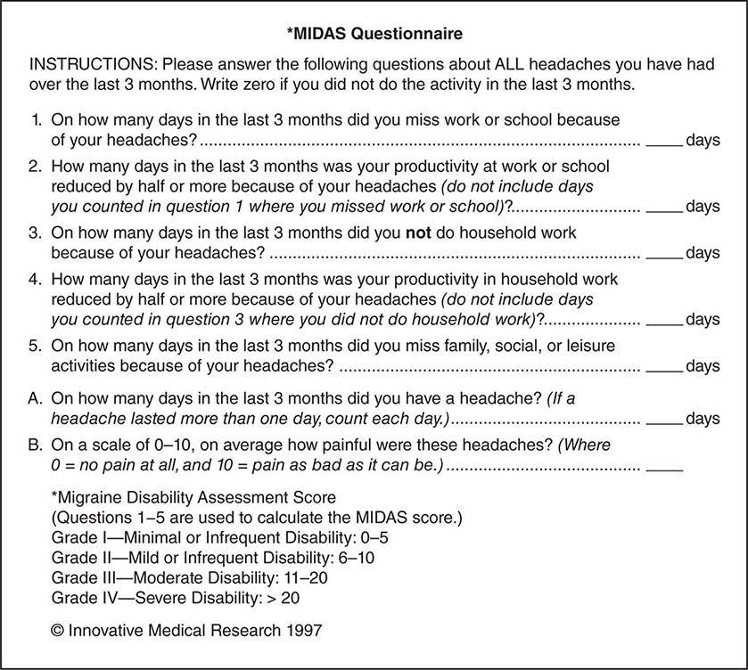
FIGURE 447-4 The Migraine Disability Assessment Score (MIDAS) Questionnaire.
Patient education is an important aspect of migraine management. Information for patients is available at sites such as www.achenet.org, the website of the American Council for Headache Education (ACHE). It is helpful for patients to understand that migraine is an inherited tendency to headache; that migraine can be modified and controlled by lifestyle adjustments and medications, but it cannot be eradicated; and that, except in some occasions in women on oral estrogens or contraceptives, migraine is not associated with serious or life-threatening illnesses.
NONPHARMACOLOGIC MANAGEMENT
Migraine can often be managed to some degree by a variety of nonpharmacologic approaches. Most patients benefit by the identification and avoidance of specific headache triggers. A regulated lifestyle is helpful, including a healthy diet, regular exercise, regular sleep patterns, avoidance of excess caffeine and alcohol, and avoidance of acute changes in stress levels, being particularly wary of the let-down effect.
The measures that benefit a given individual should be used routinely because they provide a simple, cost-effective approach to migraine management. Patients with migraine do not encounter more stress than headache-free individuals; over-responsiveness to changes in stress appears to be the issue. Because the stresses of everyday living cannot be eliminated, lessening one’s response to stress by various techniques is helpful for many patients. These may include yoga, transcendental meditation, hypnosis, and conditioning techniques such as biofeedback. For most patients, this approach is, at best, an adjunct to pharmacotherapy. Nonpharmacologic measures are unlikely to prevent all migraine attacks. If these measures fail to prevent an attack, pharmacologic approaches are then needed to abort an attack.
ACUTE ATTACK THERAPIES FOR MIGRAINE
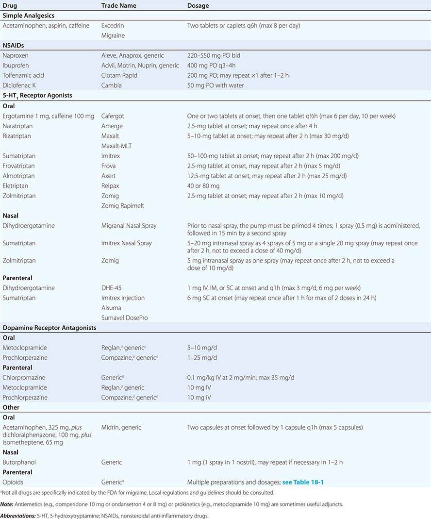
The mainstay of pharmacologic therapy is the judicious use of one or more of the many medicines that are effective in migraine (Table 447-4). The selection of the optimal regimen for a given patient depends on a number of factors, the most important of which is the severity of the attack. Mild migraine attacks can usually be managed by oral agents; the average efficacy rate is 50–70%. Severe migraine attacks may require parenteral therapy. Most drugs effective in the treatment of migraine are members of one of three major pharmacologic classes: nonsteroidal anti-inflammatory drugs, 5-HT1B/1D receptor agonists, and dopamine receptor antagonists.
TREATMENT OF ACUTE MIGRAINE |
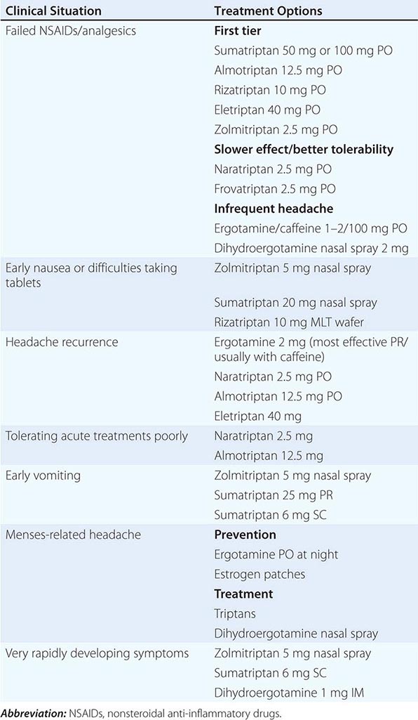
In general, an adequate dose of whichever agent is chosen should be used as soon as possible after the onset of an attack. If additional medication is required within 60 min because symptoms return or have not abated, the initial dose should be increased for subsequent attacks or a different class of drug tried as first-line treatment. Migraine therapy must be individualized; a standard approach for all patients is not possible. A therapeutic regimen may need to be constantly refined until one is identified that provides the patient with rapid, complete, and consistent relief with minimal side effects (Table 447-5).
CLINICAL STRATIFICATION OF ACUTE SPECIFIC MIGRAINE TREATMENTS |
Nonsteroidal Anti-Inflammatory Drugs (NSAIDs) Both the severity and duration of a migraine attack can be reduced significantly by NSAIDs (Table 447-4). Indeed, many undiagnosed migraineurs self-treat with nonprescription NSAIDs. A general consensus is that NSAIDs are most effective when taken early in the migraine attack. However, the effectiveness of these agents in migraine is usually less than optimal in moderate or severe migraine attacks. The combination of acetaminophen, aspirin, and caffeine has been approved for use by the U.S. Food and Drug Administration (FDA) for the treatment of mild to moderate migraine. The combination of aspirin and metoclopramide has been shown to be comparable to a single dose of oral sumatriptan. Important side effects of NSAIDs include dyspepsia and gastrointestinal irritation.
5-HT1B/1D RECEPTOR AGONISTS
Oral Stimulation of 5-HT1B/1D receptors can stop an acute migraine attack. Ergotamine and dihydroergotamine are nonselective receptor agonists, whereas the triptans are selective 5-HT1B/1D receptor agonists. A variety of triptans, 5-HT1B/1D receptor agonists—sumatriptan, almotriptan, eletriptan, frovatriptan, naratriptan, rizatriptan, and zolmitriptan—are now available for the treatment of migraine.
Each drug in the triptan class has similar pharmacologic properties but varies slightly in terms of clinical efficacy. Rizatriptan and eletriptan are the most efficacious of the triptans currently available in the United States. Sumatriptan and zolmitriptan have similar rates of efficacy as well as time to onset, with an advantage of having multiple formulations, whereas almotriptan has a similar rate of efficacy to sumatriptan and is better tolerated, and frovatriptan and naratriptan are somewhat slower in onset and are better tolerated. Clinical efficacy appears to be related more to the tmax (time to peak plasma level) than to the potency, half-life, or bioavailability. This observation is consistent with a large body of data indicating that faster-acting analgesics are more effective than slower-acting agents.
Unfortunately, monotherapy with a selective oral 5-HT1B/1D receptor agonist does not result in rapid, consistent, and complete relief of migraine in all patients. Triptans are generally not effective in migraine with aura unless given after the aura is completed and the headache initiated. Side effects are common, although often mild and transient. Moreover, 5-HT1B/1D receptor agonists are contraindicated in individuals with a history of cardiovascular and cerebrovascular disease. Recurrence of headache, within usual time course of an attack, is another important limitation of triptan use and occurs at least occasionally in most patients. Evidence from randomized controlled trials show that coadministration of a longer-acting NSAID, naproxen 500 mg, with sumatriptan will augment the initial effect of sumatriptan and, importantly, reduce rates of headache recurrence.
Ergotamine preparations offer a nonselective means of stimulating 5-HT1 receptors. A nonnauseating dose of ergotamine should be sought because a dose that provokes nausea is too high and may intensify head pain. Except for a sublingual formulation of ergotamine, oral formulations of ergotamine also contain 100 mg caffeine (theoretically to enhance ergotamine absorption and possibly to add additional analgesic activity). The average oral ergotamine dose for a migraine attack is 2 mg. Because the clinical studies demonstrating the efficacy of ergotamine in migraine predated the clinical trial methodologies used with the triptans, it is difficult to assess the clinical efficacy of ergotamine versus the triptans. In general, ergotamine appears to have a much higher incidence of nausea than triptans but less headache recurrence.
Nasal Nasal formulations of dihydroergotamine (Migranal), zolmitriptan (Zomig nasal), or sumatriptan can be useful in patients requiring a nonoral route of administration. The nasal sprays result in substantial blood levels within 30–60 min. Although in theory nasal sprays might provide faster and more effective relief of a migraine attack than oral formulations, their reported efficacy is only approximately 50–60%. Studies with a new inhalational formulation of dihydroergotamine indicate that its absorption problems can be overcome to produce rapid onset of action with good tolerability.
Parenteral Administration of drugs by injection, such as dihydroergotamine and sumatriptan, is approved by the FDA for the rapid relief of a migraine attack. Peak plasma levels of dihydroergotamine are achieved 3 min after IV dosing, 30 min after IM dosing, and 45 min after SC dosing. If an attack has not already peaked, SC or IM administration of 1 mg of dihydroergotamine suffices for about 80–90% of patients. Sumatriptan, 4–6 mg SC, is effective in ~50–80% of patients, and can now be administered by a needle-free device.
DOPAMINE RECEPTOR ANTAGONISTS
Oral Oral dopamine receptor antagonists can be considered as adjunctive therapy in migraine. Drug absorption is impaired during migraine because of reduced gastrointestinal motility. Delayed absorption occurs even in the absence of nausea and is related to the severity of the attack and not its duration. Therefore, when oral NSAIDs and/or triptan agents fail, the addition of a dopamine receptor antagonist, such as metoclopramide 10 mg or domperidone 10 mg (not available in the United States), should be considered to enhance gastric absorption. In addition, dopamine receptor antagonists decrease nausea/vomiting and restore normal gastric motility.
Parenteral Dopamine receptor antagonists (e.g., chlorpromazine, prochlorperazine, metoclopramide) by injection can also provide significant acute relief of migraine; they can be used in combination with parenteral 5-HT1B/1D receptor agonists. A common IV protocol used for the treatment of severe migraine is the administration over 2 min of a mixture of 5 mg of prochlorperazine and 0.5 mg of dihydroergotamine.
OTHER MEDICATIONS FOR ACUTE MIGRAINE
Oral The combination of acetaminophen, dichloralphenazone, and isometheptene, one to two capsules, has been classified by the FDA as “possibly” effective in the treatment of migraine. Because the clinical studies demonstrating the efficacy of this combination analgesic in migraine predated the clinical trial methodologies used with the triptans, it is difficult to compare the efficacy of this sympathomimetic compound to other agents.
Nasal A nasal preparation of butorphanol is available for the treatment of acute pain. As with all opioids, the use of nasal butorphanol has little role in migraine treatment.
Parenteral Opioids are modestly effective in the acute treatment of migraine. For example, IV meperidine (50–100 mg) is given frequently in the emergency room. This regimen “works” in the sense that the pain of migraine is eliminated. However, this regimen is clearly suboptimal for patients with recurrent headache. Opioids do not treat the underlying headache mechanism; rather, they act to alter the pain sensation, and there is evidence their use may decrease the likelihood of a response to triptans in the future. Moreover, in patients taking oral opioids, such as oxycodone or hydrocodone, habituation or addiction can greatly confuse the treatment of migraine. Opioid craving and/or withdrawal can aggravate and accentuate migraine. Therefore, it is recommended that opioid use in migraine be limited to patients with severe, but infrequent, headaches that are unresponsive to other pharmacologic approaches or who have contraindications to other therapies.
MEDICATION-OVERUSE HEADACHE
Acute attack medications, particularly opioid or barbiturate-containing compound analgesics, have a propensity to aggravate headache frequency and induce a state of refractory daily or near-daily headache called medication-overuse headache. This condition is likely not a separate headache entity but a reaction of the migraine patient to a particular medicine. Migraine patients who have two or more headache days a week should be cautioned about frequent analgesic use (see “Chronic Daily Headache” in Chap. 21).
PREVENTIVE TREATMENTS FOR MIGRAINE
Patients with an increasing frequency of migraine attacks or with attacks that are either unresponsive or poorly responsive to abortive treatments are good candidates for preventive agents. In general, a preventive medication should be considered in the subset of patients with four or more attacks a month. Significant side effects are associated with the use of many of these agents; furthermore, determination of dose can be difficult because the recommended doses have been derived for conditions other than migraine. The mechanism of action of these drugs is unclear; it seems likely that the brain sensitivity that underlies migraine is modified. Patients are usually started on a low dose of a chosen treatment; the dose is then gradually increased, up to a reasonable maximum, to achieve clinical benefit.
Drugs that have the capacity to stabilize migraine are listed in Table 447-6. Drugs must be taken daily, and there is usually a lag of between 2 to 12 weeks before an effect is seen. The drugs that have been approved by the FDA for the prophylactic treatment of migraine include propranolol, timolol, sodium valproate, topiramate, and methysergide (not available). In addition, a number of other drugs appear to display prophylactic efficacy. This group includes amitriptyline, nortriptyline, flunarizine, phenelzine, gabapentin, and cyproheptadine. Placebo-controlled trials of onabotulinum toxin type A in episodic migraine were negative, whereas, overall, placebo-controlled trials in chronic migraine were positive. Phenelzine and methysergide are usually reserved for recalcitrant cases because of their serious potential side effects. Phenelzine is a monoamine oxidase inhibitor (MAOI); therefore, tyramine-containing foods, decongestants, and meperidine are contraindicated. Methysergide may cause retroperitoneal or cardiac valvular fibrosis when it is used for >6 months, and thus monitoring is required for patients using this drug; the risk of fibrosis is about 1:1500 and is likely to reverse after the drug is stopped.
PREVENTIVE TREATMENTS IN MIGRAINEa |
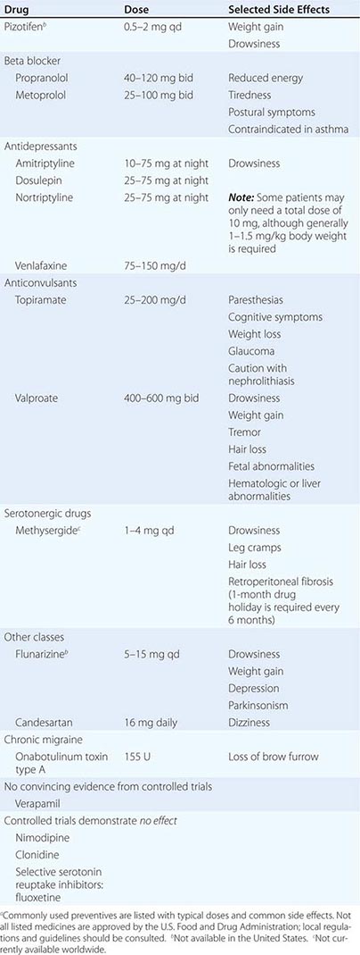
The probability of success with any one of the antimigraine drugs is 50–75%. Many patients are managed adequately with low-dose amitriptyline, propranolol, candesartan, topiramate, or valproate. If these agents fail or lead to unacceptable side effects, second-line agents such as methysergide or phenelzine can be used. Once effective stabilization is achieved, the drug is continued for ~6 months and then slowly tapered to assess the continued need. Many patients are able to discontinue medication and experience fewer and milder attacks for long periods, suggesting that these drugs may alter the natural history of migraine.
TENSION-TYPE HEADACHE
Clinical Features The term tension-type headache (TTH) is commonly used to describe a chronic head-pain syndrome characterized by bilateral tight, band-like discomfort. The pain typically builds slowly, fluctuates in severity, and may persist more or less continuously for many days. The headache may be episodic or chronic (present >15 days per month).
A useful clinical approach is to diagnose TTH in patients whose headaches are completely without accompanying features such as nausea, vomiting, photophobia, phonophobia, osmophobia, throbbing, and aggravation with movement. Such an approach neatly separates migraine, which has one or more of these features and is the main differential diagnosis, from TTH. The International Headache Society’s main definition of TTH allows an admixture of nausea, photophobia, or phonophobia in various combinations, although the appendix definition does not; this illustrates the difficulty in distinguishing these two clinical entities. In clinical practice, dichotomizing patients on the basis of the presence of associated features (migraine) and the absence of associated features (TTH) is highly recommended. Indeed patients whose headaches fit the TTH phenotype and who have migraine at other times, along with a family history of migraine, migrainous illnesses of childhood, or typical migraine triggers to their migraine attacks, may be biologically different from those who have TTH headache with none of the features. TTH may be infrequent (episodic) or occur on 15 days or more a month (chronic).
Pathophysiology The pathophysiology of TTH is incompletely understood. It seems likely that TTH is due to a primary disorder of central nervous system pain modulation alone, unlike migraine, which involves a more generalized disturbance of sensory modulation. Data suggest a genetic contribution to TTH, but this may not be a valid finding: given the current diagnostic criteria, the studies undoubtedly included many migraine patients. The name tension-type headache implies that pain is a product of nervous tension, but there is no clear evidence for tension as an etiology. Muscle contraction has been considered to be a feature that distinguishes TTH from migraine, but there appear to be no differences in contraction between the two headache types.
TRIGEMINAL AUTONOMIC CEPHALALGIAS, INCLUDING CLUSTER HEADACHE
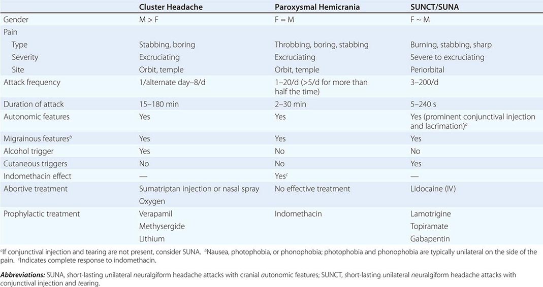
The trigeminal autonomic cephalalgias (TACs) describe a grouping of primary headaches including cluster headache, paroxysmal hemicrania, SUNCT (short-lasting unilateral neuralgiform headache attacks with conjunctival injection and tearing)/SUNA (short-lasting unilateral neuralgiform headache attacks with cranial autonomic symptoms), and hemicrania continua (Table 447-1). TACs are characterized by relatively short-lasting attacks of head pain associated with cranial autonomic symptoms, such as lacrimation, conjunctival injection, or nasal congestion (Table 447-7). Pain is usually severe and may occur more than once a day. Because of the associated nasal congestion or rhinorrhea, patients are often misdiagnosed with “sinus headache” and treated with decongestants, which are ineffective.
CLINICAL FEATURES OF THE TRIGEMINAL AUTONOMIC CEPHALALGIAS |
TACs must be differentiated from short-lasting headaches that do not have prominent cranial autonomic syndromes, notably trigeminal neuralgia, primary stabbing headache, and hypnic headache. The cycling pattern and length, frequency, and timing of attacks are useful in classifying patients. Patients with TACs should undergo pituitary imaging and pituitary function tests because there is an excess of TAC presentations in patients with pituitary tumor–related headache.
Cluster Headache Cluster headache is a relatively rare form of primary headache with a population frequency of approximately 0.1%. The pain is deep, usually retroorbital, often excruciating in intensity, nonfluctuating, and explosive in quality. A core feature of cluster headache is periodicity. At least one of the daily attacks of pain recurs at about the same hour each day for the duration of a cluster bout. The typical cluster headache patient has daily bouts of one to two attacks of relatively short-duration unilateral pain for 8 to 10 weeks a year; this is usually followed by a pain-free interval that averages a little less than 1 year. Cluster headache is characterized as chronic when there is less than 1 month of sustained remission without treatment. Patients are generally perfectly well between episodes. Onset is nocturnal in about 50% of patients, and men are affected three times more often than women. Patients with cluster headache tend to move about during attacks, pacing, rocking, or rubbing their head for relief; some may even become aggressive during attacks. This is in sharp contrast to patients with migraine, who prefer to remain motionless during attacks.
Cluster headache is associated with ipsilateral symptoms of cranial parasympathetic autonomic activation: conjunctival injection or lacrimation, rhinorrhea or nasal congestion, or cranial sympathetic dysfunction such as ptosis. The sympathetic deficit is peripheral and likely to be due to parasympathetic activation with injury to ascending sympathetic fibers surrounding a dilated carotid artery as it passes into the cranial cavity. When present, photophobia and phonophobia are far more likely to be unilateral and on the same side of the pain, rather than bilateral, as is seen in migraine. This phenomenon of unilateral photophobia/phonophobia is characteristic of TACs. Cluster headache is likely to be a disorder involving central pacemaker neurons in the posterior hypothalamic region (Fig. 447-3).
TREATMENT | CLUSTER HEADACHE |
The most satisfactory treatment is the administration of drugs to prevent cluster attacks until the bout is over. However, treatment of acute attacks is required for all cluster headache patients at some time.
ACUTE ATTACK TREATMENT
Cluster headache attacks peak rapidly, and thus a treatment with quick onset is required. Many patients with acute cluster headache respond very well to oxygen inhalation. This should be given as 100% oxygen at 10–12 L/min for 15–20 min. It appears that high flow and high oxygen content are important. Sumatriptan 6 mg SC is rapid in onset and will usually shorten an attack to 10–15 min; there is no evidence of tachyphylaxis. Sumatriptan (20 mg) and zolmitriptan (5 mg) nasal sprays are both effective in acute cluster headache, offering a useful option for patients who may not wish to self-inject daily. Oral sumatriptan is not effective for prevention or for acute treatment of cluster headache.
PREVENTIVE TREATMENTS (TABLE 447-8)
PREVENTIVE MANAGEMENT OF CLUSTER HEADACHE |
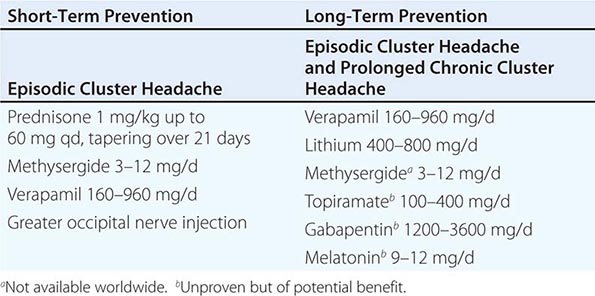
The choice of a preventive treatment in cluster headache depends in part on the length of the bout. Patients with long bouts or those with chronic cluster headache require medicines that are safe when taken for long periods. For patients with relatively short bouts, limited courses of oral glucocorticoids or methysergide (not available in the United States) can be very useful. A 10-day course of prednisone, beginning at 60 mg daily for 7 days and followed by a rapid taper, may interrupt the pain bout for many patients. Lithium (400–800 mg/d) appears to be particularly useful for the chronic form of the disorder.
Many experts favor verapamil as the first-line preventive treatment for patients with chronic cluster headache or prolonged bouts. While verapamil compares favorably with lithium in practice, some patients require verapamil doses far in excess of those administered for cardiac disorders. The initial dose range is 40–80 mg twice daily; effective doses may be as high as 960 mg/d. Side effects such as constipation and leg swelling can be problematic. Of paramount concern, however, is the cardiovascular safety of verapamil, particularly at high doses. Verapamil can cause heart block by slowing conduction in the atrioventricular node, a condition that can be monitored by following the PR interval on a standard electrocardiogram (ECG). Approximately 20% of patients treated with verapamil develop ECG abnormalities, which can be observed with doses as low as 240 mg/d; these abnormalities can worsen over time in patients on stable doses. A baseline ECG is recommended for all patients. The ECG is repeated 10 days after a dose change in patients whose dose is being increased above 240 mg daily. Dose increases are usually made in 80-mg increments. For patients on long-term verapamil, ECG monitoring every 6 months is advised.
NEUROSTIMULATION THERAPY
When medical therapies fail in chronic cluster headache, neurostimulation strategies can be used. Deep-brain stimulation of the region of the posterior hypothalamic gray matter has proven successful in a substantial proportion of patients, although its risk-benefit ratio makes it inappropriate with so many other options now available. Favorable results have also been reported with the less-invasive approach of occipital nerve stimulation, with sphenopalatine ganglion stimulation and with a noninvasive vagal nerve stimulator.
PAROXYSMAL HEMICRANIA
Paroxysmal hemicrania (PH) is characterized by frequent unilateral, severe, short-lasting episodes of headache. Like cluster headache, the pain tends to be retroorbital but may be experienced all over the head and is associated with autonomic phenomena such as lacrimation and nasal congestion. Patients with remissions are said to have episodic PH, whereas those with the nonremitting form are said to have chronic PH. The essential features of PH are unilateral, very severe pain; short-lasting attacks (2–45 min); very frequent attacks (usually more than five a day); marked autonomic features ipsilateral to the pain; rapid course (<72 h); and excellent response to indomethacin. In contrast to cluster headache, which predominantly affects males, the male-to-female ratio in PH is close to 1:1.
Indomethacin (25–75 mg tid), which can completely suppress attacks of PH, is the treatment of choice. Although therapy may be complicated by indomethacin-induced gastrointestinal side effects, currently there are no consistently effective alternatives. Topiramate is helpful in some cases. Piroxicam has been used, although it is not as effective as indomethacin. Verapamil, an effective treatment for cluster headache, does not appear to be useful for PH. In occasional patients, PH can coexist with trigeminal neuralgia (PH-tic syndrome); similar to cluster-tic syndrome, each component may require separate treatment.
Secondary PH has been reported with lesions in the region of the sella turcica, including arteriovenous malformation, cavernous sinus meningioma, pituitary pathology and epidermoid tumors. Secondary PH is more likely if the patient requires high doses (>200 mg/d) of indomethacin. In patients with apparent bilateral PH, raised cerebrospinal fluid (CSF) pressure should be suspected. It is important to note that indomethacin reduces CSF pressure. When a diagnosis of PH is considered, magnetic resonance imaging (MRI) is indicated to exclude a pituitary lesion.
SUNCT/SUNA
SUNCT (short-lasting unilateral neuralgiform headache attacks with conjunctival injection and tearing) is a rare primary headache syndrome characterized by severe, unilateral orbital or temporal pain that is stabbing or throbbing in quality. Diagnosis requires at least 20 attacks, lasting for 5–240 s; ipsilateral conjunctival injection and lacrimation should be present. In some patients, conjunctival injection or lacrimation is missing, and the diagnosis of SUNA (short-lasting unilateral neuralgiform headache attacks with cranial autonomic symptoms) can be made.
DIAGNOSIS The pain of SUNCT/SUNA is unilateral and may be located anywhere in the head. Three basic patterns can be seen: single stabs, which are usually short-lived; groups of stabs; or a longer attack comprising many stabs between which the pain does not completely resolve, thus giving a “saw-tooth” phenomenon with attacks lasting many minutes. Each pattern may be seen in the context of an underlying continuous head pain. Characteristics that lead to a suspected diagnosis of SUNCT are the cutaneous (or other) triggers of attacks, a lack of refractory period to triggering between attacks, and the lack of a response to indomethacin. Apart from trigeminal sensory disturbance, the neurologic examination is normal in primary SUNCT.
The diagnosis of SUNCT/SUNA is often confused with trigeminal neuralgia (TN) particularly in first-division TN (Chap. 455). Minimal or no cranial autonomic symptoms and a clear refractory period to triggering indicate a diagnosis of TN.
SECONDARY (SYMPTOMATIC) SUNCT SUNCT can be seen with posterior fossa or pituitary lesions. All patients with SUNCT/SUNA should be evaluated with pituitary function tests and a brain MRI with pituitary views.
Hemicrania Continua The essential features of hemicrania continua are moderate and continuous unilateral pain associated with fluctuations of severe pain; complete resolution of pain with indomethacin; and exacerbations that may be associated with autonomic features, including conjunctival injection, lacrimation, and photophobia on the affected side. The age of onset ranges from 11 to 58 years; women are affected twice as often as men. The cause is unknown.
TREATMENT | HEMICRANIA CONTINUA |
Treatment consists of indomethacin; other NSAIDs appear to be of little or no benefit. The IM injection of 100 mg of indomethacin has been proposed as a diagnostic tool, and administration with a placebo injection in a blinded fashion can be very useful diagnostically. Alternatively, a trial of oral indomethacin, starting with 25 mg tid, then 50 mg tid, and then 75 mg tid, can be given. Up to 2 weeks at the maximal dose may be necessary to assess whether a dose has a useful effect. Topiramate can be helpful in some patients. Occipital nerve stimulation probably has a role in patients with hemicrania continua who are unable to tolerate indomethacin.
Stay updated, free articles. Join our Telegram channel

Full access? Get Clinical Tree


