QUESTION 41.2
A. Diffuse extracapillary proliferative glomerulonephritis
B. Diffuse intracapillary proliferative glomerulonephritis
C. Focal glomerulonephritis
D. Membranous glomerulopathy
E. Normal glomerular morphology
3. A renal biopsy from a patient with a severe infection reveals a diffuse intracapillary proliferation with leukocytic infiltration and obliteration of capillary lumina. Subepithelial electron-dense “humps” are found on EM. Which of the following IF findings would be consistent with an infection caused by Staphylococcus aureus?
A. Diffuse capillary loop deposits of C3
B. Diffuse mesangial IgM deposits
C. Linear deposition of IgG and C3 along GBM
D. Granular mesangial and glomerular capillary IgG and C3 deposits
E. Granular mesangial and glomerular capillary IgA deposits
4. Which of the following morphologic or IF features favors a diagnosis of C3 glomerulopathy over that of classic membranoproliferative glomerulonephritis (MPGN)?
A. Mesangial interposition with GBM duplication
B. Mesangial proliferation
C. Mesangial and subendothelial deposits of C3 and IgG
D. Mesangial, subendothelial, and intramembranous deposits of C3 and IgG
E. Mesangial and intramembranous deposits of C3 alone
5. A 12-year-old boy has nephrotic syndrome and low complement levels. The most significant findings on renal biopsy are demonstrated in these PAS (A) and methenamine silver–stained sections (B). Mesangial and glomerular capillary deposition of C3 and IgG was detected by IF. EM showed large subendothelial electron-dense deposits with mesangial interposition. Which of the following is the most likely diagnosis?
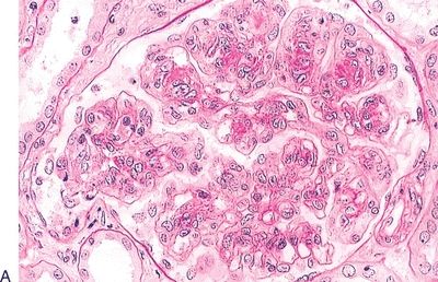
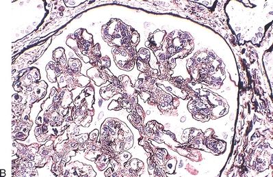
QUESTION 41.5
A. Dense deposit disease (MPGN, type II)
B. MPGN, type I
C. Membranous glomerulopathy
D. Minimal change glomerulopathy
E. Postinfectious glomerulonephritis
A. Dense deposit disease
B. Light chain disease
C. Membranoproliferative (mesangiocapillary) glomerulonephritis, type I
D. Membranous glomerulopathy
E. Minimal change glomerulopathy
7. Which of the following is true about crescentic glomerulonephritis?
A. Crescents consist exclusively of parietal cells of the Bowman capsule
B. Finding 20% of glomeruli with crescents supports the diagnosis
C. Most cases are associated with a known underlying condition
D. Most cases are associated with linear deposition of anti-GBM IgG
E. Prognosis depends on extent of crescent formation and IF pattern
8. Which of the following glomerular lesions is shown in this picture of a PAS-stained section?
QUESTION 41.8
A. Cellular crescent
B. Fibrocellular crescent
C. Fibrous crescent
D. Obsolescent glomerulus
E. Segmental scarring
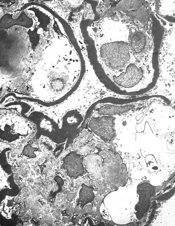
9. A 4-year-old child presents with nephrotic syndrome, and a renal biopsy shows no light microscopic changes. Ultrastructural changes are shown in this picture. Which of the following immunofluorescence findings would be most likely present?
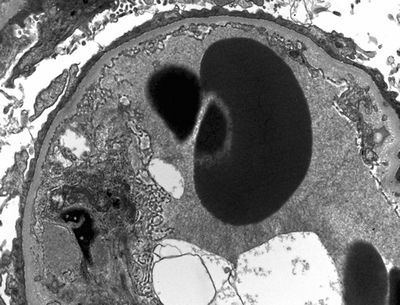
QUESTION 41.9
A. Absence of C3 and Ig deposits
B. Continuous granular C3 deposition along capillary wall
C. Granular IgG/C3 deposits along GBM
D. Linear IgG deposition along GBM
E. Mesangial IgA deposits
10. A clinical evaluation of a young adult with generalized edema shows heavy proteinuria (4.5 g/day). The past medical history is unremarkable. In the United States, which of the following is the most common glomerular disease accounting for this presentation?
A. Diabetic nephropathy
B. Focal segmental glomerulosclerosis
C. IgA nephropathy (Berger disease)
D. Membranous glomerulonephritis
E. Minimal change disease
11. An adult patient presents with nephrotic syndrome, and a renal biopsy reveals the glomerular changes depicted in this photomicrograph. Which of the following ultrastructural alterations is most likely found in the glomeruli that appear normal on light microscopy?
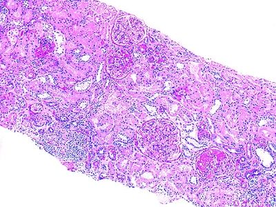
QUESTION 41.11
A. Effacement of epithelial cell foot processes
B. Mesangial interposition
C. Subendothelial deposits
D. Subepithelial humps
E. Subepithelial deposits with spikes of GBM material
12. Focal segmental glomerulosclerosis (FSGS) is the usual morphologic pattern of HIV-associated nephropathy (HIVAN). Which of the following light microscopic changes is most characteristic of HIVAN?
A. Argyrophilic spikes on the basement membrane
B. Crescent formation
C. Glomerular collapse and tubular interstitial fibrosis
D. Obsolescent glomeruli
E. None of the above
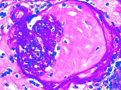
13. A renal biopsy is performed on a 34-year-old man for the evaluation of proteinuria in the nephrotic range. A representative silver-stained section is shown in the photomicrograph. IF demonstrates finely granular deposits of IgG and C3 along the capillary wall. EM shows effacement of foot processes, associated with numerous subepithelial electron-dense deposits, which are separated from each other by extensions of the basement membrane. Which of the following is the most likely diagnosis?
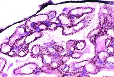
QUESTION 41.13
A. Dense deposit disease
B. Focal segmental glomerulosclerosis
C. Membranoproliferative glomerulonephritis
D. Membranous glomerulonephritis
E. Minimal change glomerulopathy
14. A previously healthy 35-year-old woman presents with nephrotic syndrome. She has no other known significant condition. Laboratory tests reveal circulating IgG4 directed against the phospholipase A2 receptor (PLA2R). Which of the following is the most likely glomerular disease?
A. Dense deposit disease
B. Focal segmental glomerulosclerosis
C. Membranoproliferative glomerulonephritis
D. Membranous glomerulonephritis
E. Minimal change nephrotic syndrome
15. A 55-year-old woman with a 15-year history of type 2 diabetes mellitus is found to have microalbuminuria. All other renal function tests are normal. At this early stage, the most likely glomerular change of diabetic nephropathy is:
A. Expansion of the mesangial matrix
B. Fibroepithelial crescents
C. Insudative lesions such as fibrin caps and capsular drops
D. Mesangial intercapillary nodules (of Kimmelstiel-Wilson)
E. Thickening of the basement membrane
16. The glomerular lesion seen in this photomicrograph was found in a renal biopsy from a patient with long-standing diabetes mellitus. How is this glomerular change called?

QUESTION 41.16
A. Capsular drop
B. Diffuse diabetic glomerulosclerosis
C. Fibrin cap
D. Microaneurysm
E. Nodular diabetic glomerulosclerosis
17. Which of the following is the most common form of glomerulonephritis worldwide and manifests with recurrent gross or microhematuria?
A. Alport syndrome
B. IgA nephropathy (Berger disease)
C. Pauci-immune crescentic glomerulonephritis
D. Postinfectious glomerulonephritis
E. Thin membrane disease
18. Microscopic hematuria is discovered in an otherwise healthy 20-year-old man. There is a family history of similar abnormality. A renal biopsy appears normal on light microscopy, but the glomerular basement membrane is less than 200 nm thick on EM. Which of the following extrarenal abnormalities is most likely associated with this condition?
A. Deafness
B. Hypoplasia of patella
C. Lens dislocation
D. Nail dysplasia
E. None of the above
19. A hereditary form of glomerulopathy caused by defective collagen synthesis and associated with sensorineural deafness and ocular abnormalities is:
A. Alport syndrome
B. Berger disease
C. Fabry disease
D. Onychoosteodysplasia
E. Thin membrane disease
20. Which of the following conditions is most likely to be associated with the type of “bloodless” glomerulus, as depicted in this photomicrograph?
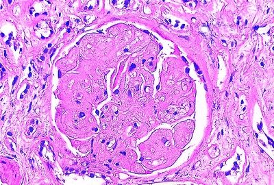
QUESTION 41.20
A. Cryoglobulinemia
B. Hemolytic uremic syndrome
C. Multiple myeloma
D. Streptococcal infection
E. Systemic lupus erythematosus
21. A 35-year-old man presents with nephritic syndrome. Several years prior to this, he suffered from an episode of generalized purpura due to cryoglobulinemia. Which of the following ultrastructural changes is most characteristic of glomerular disease secondary to cryoglobulinemia?
A. Diffuse vacuolization of glomerular endothelial cells
B. Laminated intraepithelial inclusions called “zebra bodies”
C. Subendothelial deposits with a fingerprint-like pattern
D. Subepithelial electron-dense “humps”
E. Tubulovesicular inclusions in endothelial cells
22. A 60-year-old woman with a long history of rheumatoid arthritis comes to medical attention because of heavy proteinuria. Which of the following alterations is most likely to be identified on a renal biopsy?
A. Amyloid deposition
B. IgA mesangial deposition
C. Immune complex deposition
D. Light chain mesangial deposition
E. Urate crystal deposition within tubules
23. A 40-year-old man presents with proteinuria in the nephrotic range, microhematuria, and hypertension. The patient does not have monoclonal gammopathy, lymphoproliferative disorders, or cryoglobulinemia. A renal biopsy reveals expansion of the mesangium and thickening of the glomerular capillary wall. IF staining with antibodies to IgG is shown in this picture. A Congo red stain is negative. Which of the following type of glomerular deposits would be most likely found by EM?
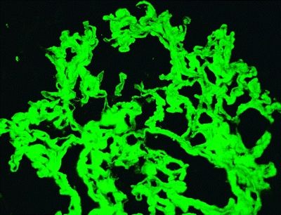
QUESTION 41.23
A. Microtubular structures with a 20 to 50 nm diameter
B. Randomly arranged, rigid 8- to 10-nm fibrils
C. Randomly arranged, rigid 18- to 24-nm fibrils
24. A 30-year-old woman with systemic lupus erythematosus (SLE) undergoes a renal biopsy because of proteinuria and microhematuria. The biopsy shows glomerular changes consistent with class IV lupus nephritis according to the new classification by the International Society of Nephrology and the Renal Pathology Society (ISN/RPS). Which of the following glomerular changes is present in this renal biopsy?
A. Minimal mesangial lupus nephritis
B. Mesangial proliferative lupus nephritis
C. Focal lupus nephritis
D. Diffuse lupus nephritis
E. Membranous lupus nephritis
F. Advanced sclerosing lupus nephritis
25. Which of the following morphologic parameters is used in the evaluation of the activity of lupus nephritis?
A. Cellular crescents
B. Fibrous crescents
C. Glomerulosclerosis
D. Interstitial fibrosis
E. Tubular atrophy
26. A 65-year-old man with heavy proteinuria and hypertension undergoes a renal biopsy. The main biopsy finding consists of diffuse and nodular mesangial accumulation of PAS-positive material. IF reveals deposition of kappa light chain throughout the mesangium and along the GBM, as shown in this picture. On light microscopy, the glomerular changes of this condition may be very similar to

QUESTION 41.26


