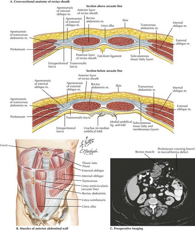
Abdominal Wall Anatomy and Ostomy Sites

Chapter 20


Abdominal Wall Anatomy and Ostomy Sites


Surgical Principles


The general location of the stoma will be determined by the type of ostomy required (proximal or distal bowel, small or large intestine) and by the reach of the mesentery associated with the bowel to be brought through the skin. Ideally, the patient should be examined preoperatively and potential ostomy sites marked by the surgeon or a trained enterostomal nurse. In cases in which the exact stoma location is unknown preoperatively, multiple acceptable locations may be marked to provide the surgeon with several options for placement. Markings are usually performed with permanent marker and may be scored with a needle intraoperatively to avoid removal during surgery.


The planned ostomy site should be placed over the rectus abdominis muscle whenever possible. Care should be taken to observe the patient standing, sitting, and reclining to identify and avoid skin creases and bony prominences that may cause pouching difficulties. Placement of the site should avoid the patient’s belt line and ensure easy visualization and reach by the patient, accounting for any functional limitations. Ideally, stomas should be located far enough from incisions to allow for placement of a stoma appliance lateral to existing or new incisions. By locating the stoma in the upper abdomen in obese patients, the surgeon may avoid placing the stoma through the pannus, decrease the amount of subcutaneous tissue between fascia and skin, and provide better visualization of the stoma for the patient.


Abdominal Wall Anatomy


The fasciae of the external and internal oblique and transversus abdominis muscles converge at the linea semilunaris, lateral to the rectus muscles (Fig. 20-1). Medially, these fasciae converge again at the linea alba. Superior to the linea semicircularis (arcuate line), usually at the umbilicus, the fasciae divide to encircle the rectus muscles completely, forming an anterior and a posterior rectus sheath. Inferior to the linea semicircularis, all layers of the fasciae are anterior to the rectus muscles, with only the transversalis fascia posterior.


Stay updated, free articles. Join our Telegram channel

Aug 12, 2016 | Posted by in ANATOMY | Comments Off on Abdominal Wall Anatomy and Ostomy Sites

Full access? Get Clinical Tree

Get Clinical Tree app for offline access