FIGURE 25–1. Group A streptococcus (GAS) Gram stain. Note the oval cocci chaining end to end (arrow). (Image contributed by professor Shirley Lowe, University of California, San Francisco School of Medicine, with permission.)
Oval cells arranged in chains end to end
Streptococci grow best in enriched media under aerobic or anaerobic conditions (facultative). Sheep blood agar is preferred because it satisfies the growth requirements and also serves as an indicator for patterns of hemolysis. The colonies are small, ranging from pinpoint size to 2 mm in diameter, and they may be surrounded by a zone where the erythrocytes suspended in agar have been hemolyzed. When the zone is clear, this state is called β-hemolysis (Figure 25–2). When the zone is hazy with a green discoloration of the agar, it is called α-hemolysis. Streptococci are metabolically active, attacking a variety of carbohydrates, proteins, and amino acids. Glucose fermentation yields mostly lactic acid. In contrast to staphylococci, streptococci are catalase-negative.
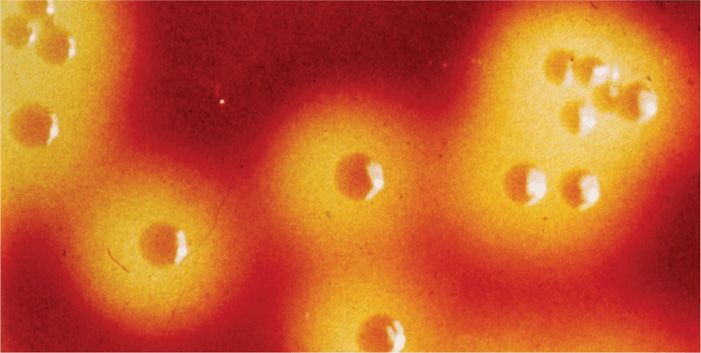
FIGURE 25–2. β-Hemolysis. Colonies of group A streptococci (GAS) on sheep blood agar plates are surrounded by a zone of complete clearing of the RBCs suspended in the agar. (Reproduced with permission from Nester EW: Microbiology: A Human Perspective, 6th edition. 2009.)
β-Hemolysis is clear
α-Hemolysis shows greening of blood agar
Catalase-negative
CLASSIFICATION
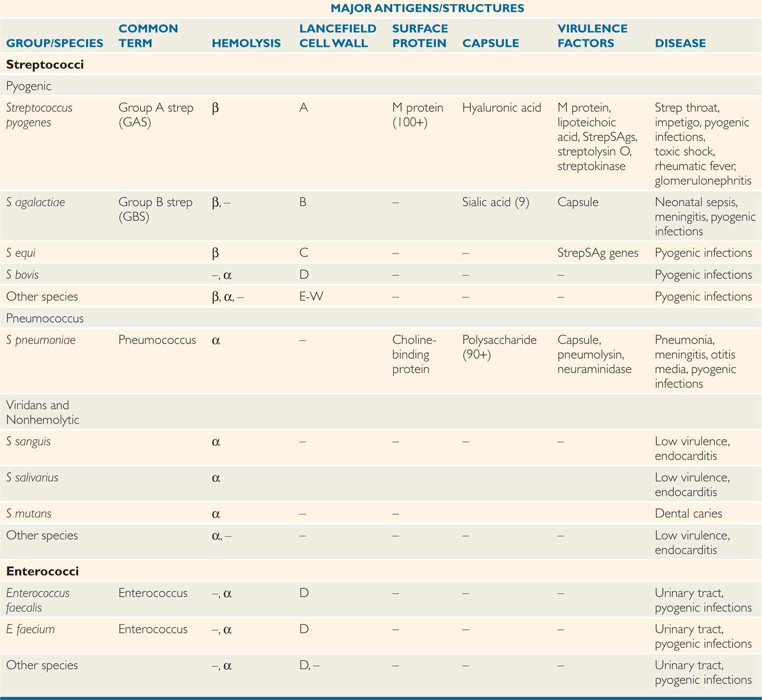
At the turn of the 20th century, a classification based on hemolysis and biochemical tests was sufficient to associate some streptococcal species with infections in humans and animals. Rebecca Lancefield, who demonstrated carbohydrate antigens in cell wall extracts of the β-hemolytic streptococci, put this taxonomy on a sounder basis. Her studies formed a classification by serogroups (eg, A, B, C), each of which is generally correlated with one of the previously established species. Later it was discovered that some nonhemolytic streptococci had the same cell wall antigens. Over the years it has become clear that possession of one of the Lancefield antigens defines a particularly virulent segment of the streptococcal genus regardless of hemolytic patterns. These are called the pyogenic streptococci, and in medical circles they are now better known by their Lancefield letter than the older species name. Pediatricians instantly recognize GBS as an acronym for group B streptococcus, but may be confused by use of the proper name, Streptococcus agalactiae (Table 25–1).
TABLE 25–1 Classification of Streptococci and Enterococci
Lancefield antigens are cell wall carbohydrates
Presence of Lancefield antigens defines the pyogenic streptococci
For practical purposes, the type of hemolysis and certain biochemical reactions remain valuable for the initial recognition and presumptive classification of streptococci, and as an indication of what subsequent taxonomic tests to perform. Thus, β-hemolysis indicates that the strain has one of the Lancefield group antigens, but some Lancefield-positive strains or groups may be α-hemolytic or even nonhemolytic. The streptococci are considered here as follows: (1) pyogenic streptococci (Lancefield groups); (2) pneumococci; and (3) viridans and other streptococci (Table 25–1).
Hemolysis is a practical guide to classification
Only pyogenic streptococci are P-hemolytic
 Pyogenic Streptococci
Of the many Lancefield groups, the ones most frequently isolated from humans are A, B, C, F, and G. Of these, groups A (S pyogenes) and B (S agalactiae) are the most common causes of serious disease. The group D carbohydrate is found in the genus Enterococcus, which used to be classified among the streptococci.
Groups A and B streptococci are the most common causes of disease
 Pneumococci
This category contains a single species, S pneumoniae, commonly called the pneumococcus. Its distinctive feature is the presence of a capsule composed of polysaccharide polymers that vary in antigenic specificity. More than 90 capsular immunotypes have been defined. Although the pneumococcal cell wall shares some common antigens with other streptococci, it does not possess any of the Lancefield group antigens. Streptococcus pneumoniae is α-hemolytic.
Pneumococci have an antigenic polysaccharide capsule
 Viridans and Other Streptococci
Viridans streptococci are α-hemolytic and lack both the group carbohydrate antigens of the pyogenic streptococci and the capsular polysaccharides of the pneumococcus. The term encompasses several species, including S salivarius and S mitis. Viridans streptococci are members of the resident oral flora of humans. They rarely demonstrate invasive qualities. A variety of other streptococci may be encountered, which lack the features of the pyogenic streptococci or pneumococci; these would be classified with the viridans group, except that they are not α-hemolytic. Such strains are usually assigned descriptive terms such as non-hemolytic streptococci or microaerophilic streptococci. They have been less thoroughly studied, but generally have the same biologic behavior as the viridans streptococci.
Viridans and nonhemolytic species lack Lancefield antigens or capsules
Group A Streptococci (Streptococcus pyogenes)
 BACTERIOLOGY
MORPHOLOGY AND GROWTH
Group A streptococci (GAS) typically appear in purulent lesions or broth cultures as spherical or ovoid cells in chains of short to medium length (4-10 cells). On blood agar plates, colonies are usually compact, small, and surrounded by a 2 to 3 mm zone of β-hemolysis (Figure 25–2), which is easily seen and sharply demarcated. β-Hemolysis is caused by either of two hemolysins, streptolysin S and the oxygen-labile streptolysin O, both of which are produced by most group A strains. Strains that lack streptolysin S are β-hemolytic only under anaerobic conditions, because the remaining streptolysin O is not active in the presence of oxygen. This feature is of practical importance, because such strains would be missed in clinical laboratories if cultures were incubated only aerobically.
Streptolysin O or S cause β-hemolysis
Aerobically, only S is active
STRUCTURE
The structure of GAS is illustrated in Figure 25–3. The cell wall is built on a peptidoglycan matrix that provides rigidity, as in other Gram-positive bacteria. Within this matrix lies the group carbohydrate antigen, which by definition is present in all GAS. A number of other molecules such as M protein and lipoteichoic acid (LTA) are attached to the cell wall, but extend beyond, often in association with, the hair-like pili. Group A streptococci are divided into more than 100 serotypes based on antigenic differences in the M protein.
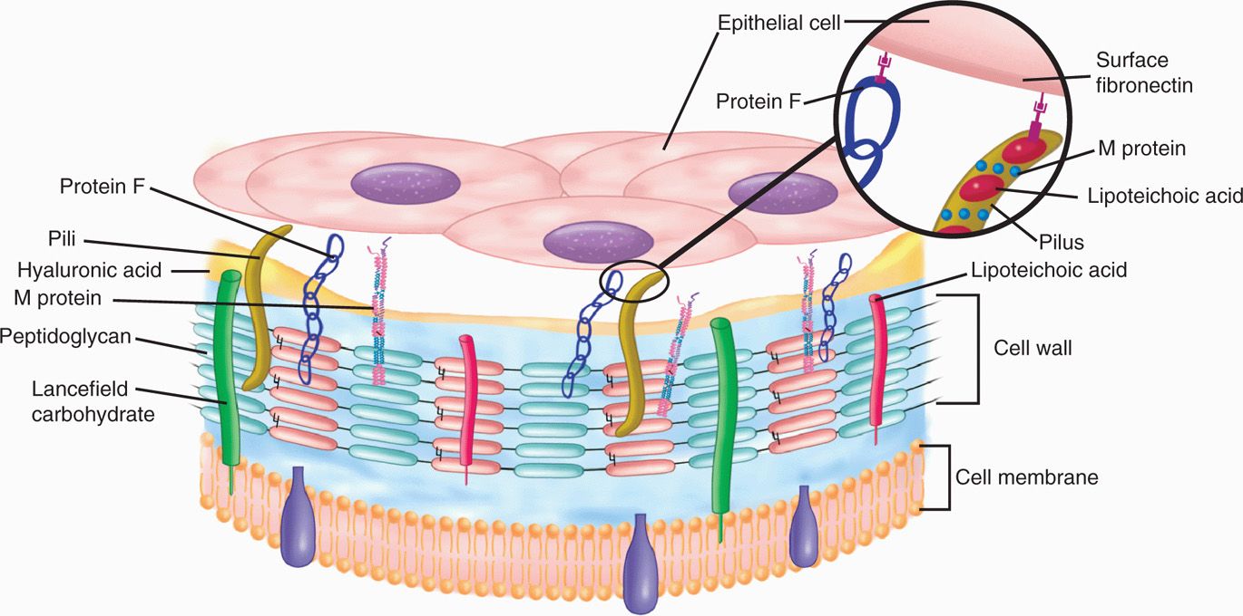
FIGURE 25–3. Antigenic structure of GAS and adhesion to an epithelial cell. The location of peptidoglycan and Lancefield carbohydrate antigens in the cell wall is shown in the diagram. M protein and lipoteichoic acid are associated with the cell surface and the pili. Lipoteichoic acid and protein F mediate binding to fibronectin on the host surface.
Wall contains group antigen with multiple surface molecules extending beyond
 M Protein
The M protein itself is a fibrillar coiled-coil molecule (Figure 25–4) with structural homology to myosin. Its carboxy terminus is rooted in the peptidoglycan of the cell wall, and the amino-terminal regions extend out from the surface. The specificity of the multiple serotypes of M protein is determined by variations in the amino sequence of the amino-terminal portion of the molecule. Because of its exposed location, this part of the M protein is also the most available to immune surveillance. The middle part of the molecule is less variable, and some carboxy-terminal regions are conserved across many M types. There is increasing evidence that some of the many known biologic functions of M protein can be assigned to specific domains of the molecule. This includes both antigenicity and the capacity to bind other molecules such as fibrinogen, serum factor H, and immunoglobulins. There are more than 100 immunotypes of M protein, which are the basis of a subtyping system for GAS.
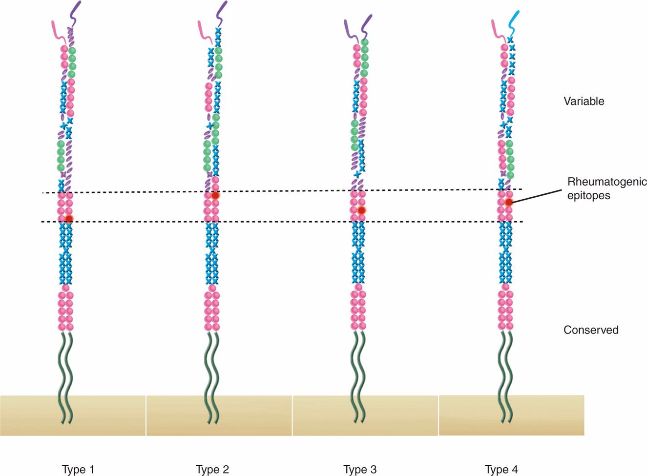
FIGURE 25–4. M protein. The coiled-coil structure of M protein is shown for four hypothetical serotypes. The most variable parts of the molecule are oriented to the outside and provide the antiphagocytic effect and serologic specificity for each type. The conserved portions are rooted in the cell wall and are homologous in structure. All four types contain epitopes that may stimulate the cross-reactive immune reactions seen in rheumatic fever.
Coiled-coil structure is similar to myosin
Antigenicity and function differ in domains of the molecule
100+ M protein serotypes exist
 Other Surface Molecules
A number of surface proteins have been described on the basis of their similarity with M protein or some unique binding capacity. Of these, a fibronectin-binding protein F and LTA are both exposed on the streptococcal surface (Figure 25–3) and play a role in pathogenesis. An IgG-binding protein has the capacity to bind the Fc portion of antibodies in much the same way as staphylococcal protein A. In principle, this could interfere with opsonization by creating a covering of antibody molecules on the streptococcal surface that are facing the “wrong way.” Many GAS have a nonantigenic hyaluronic acid capsule. Although this capsule has been shown to be antiphagocytic, its role in disease is clouded by the fact that strains which lack it are still fully virulent.
Protein F and LTA bind fibronectin
Hyaluronic acid capsule may be present
EXTRACELLULAR PRODUCTS
 Streptolysin O
Streptolysin O is a pore-forming cytotoxin, lysing leukocytes, tissue cells, and platelets. The toxin inserts directly into the cell membrane of host cells, forming transmembrane pores in a manner similar to complement and staphylococcal α-toxin. Streptolysin O is antigenic, and the quantitation of antibodies against it is the basis of a standard serologic test called antistreptolysin O (ASO).
Streptolysin O is pore-forming and antigenic
 Streptococcal Superantigen Toxins
Just as with Staphylococcus aureus, approximately 10% of GAS produce one of a family of exotoxins whose major biologic effect is through the superantigen (SAg) mechanism (Figure 22–7). Over many decades, these toxins have been assigned a number of names linked to their association with scarlet fever (erythrogenic toxin) and with streptococcal toxic shock (streptococcal pyrogenic exotoxins [Spe]). As with S aureus, there are several antigenically distinct proteins (SpeA, SpeB, and so on). Because the staphylococcal and streptococcal SAgs have similar amino acid structure and biologic activity, in this book they are called StaphSAgs and StrepSAgs. StrepSAgs have multiple effects, including fever, rash (scarlet fever), T-cell proliferation, B-lymphocyte suppression, and heightened sensitivity to endotoxin. Most of these actions are due to cytokine release through the superantigen mechanism. At least one StrepSAg (SpeB) also has direct enzymatic activity digesting tissue and extracellular matrix proteins.
StrepSAgs are produced by some strains
StrepSAgs and StaphSAgs are superantigens
 Other Extracellular Products
Most strains of GAS produce a number of other extracellular products including streptokinase, hyaluronidase, nucleases, and a C5a peptidase. The C5a peptidase is an enzyme that degrades complement component C5a, the main factor that attracts phagocytes to sites of complement deposition. The enzymatic actions of the others likely play some role in tissue injury or spread, but no specific roles have been defined. Some are antigenic and have been the basis of serologic tests. Streptokinase causes lysis of fibrin clots through conversion of plasminogen in normal plasma to the protease plasmin.
C5a peptidase degrades complement
Streptokinase converts plasminogen to plasmin
![]() GROUP A STREPTOCOCCAL DISEASE
GROUP A STREPTOCOCCAL DISEASE
EPIDEMIOLOGY
 Pharyngitis
Group A streptococci are the most common bacterial cause of pharyngitis in schoolage children 5 to 15 years of age. Transmission is person-to-person from the large droplets produced by infected persons during coughing, sneezing, or even conversation (Figure 25–5). This droplet transmission is most efficient at the short distances (2-5 feet) at which social interactions commonly take place in families and schools, particularly in fall and winter months. Asymptomatic carriers (<1%) may also be the source of GAS, particularly if colonized in the nose as well as the throat. Although GAS survive for some time in dried secretions, environmental sources and fomites are not important means of spread. Unless the condition is treated, the organisms persist for 1 to 4 weeks after symptoms have disappeared.
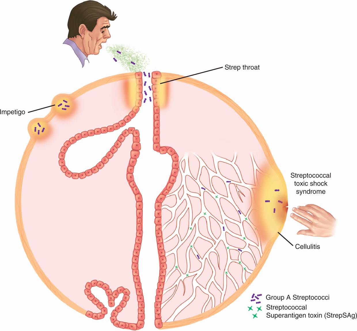
FIGURE 25–5. GAS disease overview. The primary sources of infection are respiratory droplets or direct contact with the skin. Impetigo results from minor trauma such as insect bites in skin transiently colonized with GAS. In streptococcal toxic shock, StrepSags producing GaS in a superficial lesion spread into the bloodstream. Note both toxin and bacteria are circulating.
Most common bacterial cause of sore throat
Droplets spread over short distances from throat and nasal sites
 Impetigo
Impetigo occurs when transient skin colonization with GAS is combined with minor trauma such as insect bites. The tiny skin pustules are spread locally by scratching and to others by direct contact or shared fomites such as towels. Impetigo is most common in summer months when insects bite and when the general level of hygiene is low. The M protein types of GAS most commonly associated with impetigo are different from those causing respiratory infection.
Skin colonization plus trauma leads to impetigo
 Wound and Puerperal Infections
Group A streptococci, once a leading cause of postoperative wound and puerperal infections, retain this potential, but the conditions favoring these diseases are now less common in developed countries. As with staphylococci, transmission from patient-to-patient is by the hands of physicians or other medical attendants who fail to follow recommended hand-washing practices. Organisms may be transferred from another patient or may come from the healthcare workers themselves.
Hospital outbreaks are linked to carriers
 Streptococcal Toxic Shock Syndrome
Since the late 1980s, a severe invasive form of GAS soft tissue infection appeared with increased frequency worldwide. Rapid progression to death in only a few days occurred in previously healthy persons, including Muppet creator Jim Henson of Sesame Street fame. The outstanding features of these infections are their multiorgan involvement, suggesting a toxin and rapid invasiveness with spread to the bloodstream and distant organs. Soft-tissue necrosis and streptococcal gangrenous myositis can rapidly ensure without the trauma associated with clostridial gas gangrene (Chapter 29). The toxic features together with the discovery that almost all the isolates produce StrepSAgs have caused this syndrome to be labeled streptococcal toxic shock syndrome (STSS).
STSS may be fatal in healthy persons
Strains produce StrepSAgs
 Poststreptococcal Sequelae
The association between GAS and the inflammatory disease acute rheumatic fever (ARF) is based on epidemiologic studies linking GAS pharyngitis, the clinical features of rheumatic fever, and heightened immune responses to streptococcal products. ARF does not follow skin or other nonrespiratory infection with GAS. Although some M types are more “rheumatogenic,” it is not practical to define risk in advance. The general approach is that recurrences of ARF can be triggered by infection with any GAS. Injury to the heart caused by recurrences of ARF leads to rheumatic heart disease, a major cause of heart disease worldwide. Although ARF has declined in developed countries, resurgence in the form of small regional outbreaks began in the late 1980s. These outbreaks involved children of a higher socioeconomic status than that previously associated with ARF and a shift in prevalent M types. The underlying basis of the resurgence is unknown. In contrast, ARF is rampant in many developing countries, particularly in Africa, the Middle East, India, and South America.
ARF follows respiratory, not skin, infection
Rheumatic heart disease is produced by recurrent ARF
Poststreptococcal glomerulonephritis may follow either respiratory or cutaneous GAS infection and involves only certain “nephritogenic” strains. It is more common in temperate climates where insect bites lead to impetigo. The average latent period between infection and glomerulonephritis is 10 days from a respiratory infection, but generally about 3 weeks from a skin infection. Nephritogenic strains are limited to a few M types and seem to have declined in recent years.
Glomerulonephritis follows respiratory or skin infection
Only nephritogenic strains are involved
PATHOGENESIS
 Acute Infections
As with other pathogens, adherence to mucosal surfaces is a crucial step in initiating disease (Figure 25–6). Along with pili, a dozen specific adhesins have been described that facilitate the ability of the GAS to adhere to epithelial cells of the nasopharynx and/or skin. Of these, the most important are M protein, LTA, and protein F. In the nasopharynx, all three appear to be involved in mediating attachment to the fatty acid binding sites in the glycoprotein fibronectin covering the epithelial cell surface. The role of M protein in the pharynx is not direct, but it appears to function as an anchor for LTA, which is essential for it to reach its binding site (Figure 25–3).
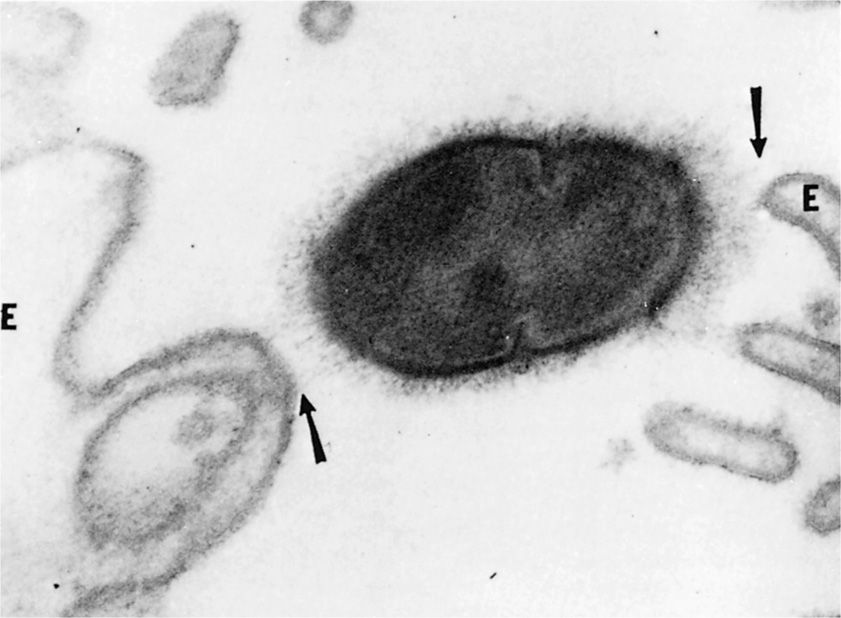
FIGURE 25–6. GAS is shown attaching to the cell membrane of a human oral epithelial cell (e). Note the hair-like pili (arrows), which mediate the attachment. as in Figure 23–3, both M protein and lipoteichoic acid are associated with the pili. (reproduced with permission from Beachey eh, Ofek I. J exp Med 1976;143:764.)
Surface molecules binding to fibronectin is important first step
M protein supports nasopharyngeal cell adherence
However, M protein appears to be direct and dominant in binding to the skin through its ability to interact with subcorneal keratinocytes, the most numerous cell type in cutaneous tissue. This adherence takes place at domains of the M protein that bind to receptors on the keratinocyte surface. Protein F is also involved primarily in adherence to antigen-presenting Langerhans cells. Expression of M protein and protein F is regulated in response to environmental conditions (O2, CO2), which could play a role in establishing the microbe or in relation to the immune response.
M protein and protein F are involved in keratinocyte binding
Expression is environmentally regulated
Clinical evidence makes it clear that GAS have the capacity to be highly invasive. The events following attachment that trigger invasion are only starting to be understood. It appears that M protein, protein F, and other fibronectin-binding proteins are required for the invasion of nonprofessional phagocytes. There is also evidence that StrepSAg genes are linked to invasiveness. The invasion itself involves integrin receptors and is accompanied by cytoskeleton rearrangements, but the molecular events do not yet make a coherent story.
Multiple factors involved in invasion
After the initial events of attachment and invasion, the concerted activity of the M protein, immunoglobulin-binding proteins, and the C5a peptidase play the key roles in allowing the streptococcal infection to continue (Figure 25–7). M protein plays an essential role in GAS resistance to phagocytosis because of the ability of domains of the molecule to bind serum factor H. This leads to a diminished availability of alternative pathway-generated complement component C3b for deposition on the streptococcal surface in the same manner as polysaccharide capsules (Figure 22–4). In the presence of M type-specific antibody, classical pathway opsonophagocytosis proceeds, and the streptococci are rapidly killed. As a second antiphagocytic mechanism, the C5a peptidase inactivates C5a and thus blocks chemotaxis of polymorphonuclear neutrophils (PMNs) and other phagocytes to the site of infection.
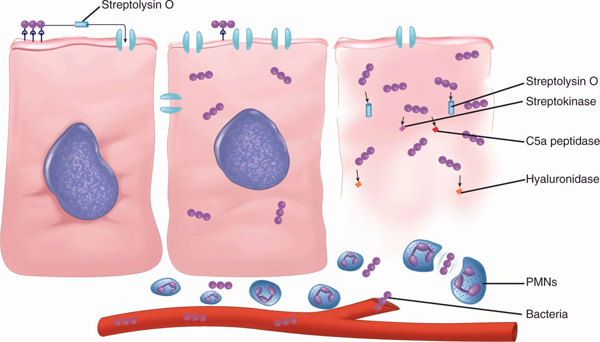
FIGURE 25–7. GAS disease, cellular view. The cellular events are similar to that of Staphylococcus aureus (see Figure 24–4). Streptolysin O is a pore-forming toxin, and there are many extracellular products. A difference is that although S aureus tends to be localized, GAS tend to spread diffusely, as shown in the cell on the right. This may be due to hyaluronidase (spreading factor) or resistance to phagocytosis. Below the cells, factor H binding is mediating GAS escaping the polymorphonuclear neutrophils (pMNs).
Antiphagocytic M protein binds factor H
Surface C3b deposition is diminished
C5a peptidase blocks phagocyte chemotaxis
The precise role of other bacterial factors in the pathogenesis of acute infection is uncertain, but the combined effect of streptokinase, DNAase, and hyaluronidase may prevent effective localization of the infection, whereas the streptolysins produce tissue injury and are toxic to phagocytic cells. Antibodies against these components are formed in the course of streptococcal infection but are not known to be protective.
Other virulence factors contribute to spread and injury
In STSS, as with staphylococcal toxic shock syndrome, the findings of shock, renal impairment, coagulopathy, and rash seem to be explained by the massive cytokine release stimulated by the superantigenicity of the StrepSAgs. Exotoxin production, however, does not explain the enhanced invasiveness of GAS, which is an added feature of STSS compared to its staphylococcal counterpart. Although the enzymatic activity of some StrepSAgs have been linked to invasiveness, the underlying mechanisms are unclear. One theory is that STSS may be due to the horizontal transfer of StrepSAg genes to GAS clones with enhanced invasive potential, a deadly combination.
Superantigenicity of StrepSAgs contributes to STSS
Invasive component is unexplained
 Poststreptococcal Sequelae
Acute Rheumatic Fever
Of the many theories advanced to explain the role of GAS in ARF, an autoimmune mechanism related to antigenic similarities between streptococci antigens and human tissues has the most experimental support. Streptococcal pharyngitis patients who develop ARF have higher levels of antistreptococcal and autoreactive antibodies than those who do not. Some of these antibodies have been shown to react with both heart tissue and streptococcal antigens.
ARF is an autoimmune state induced by streptococcal infection
The antigen stimulating these antibodies is most probably M protein, but the group A carbohydrate is also a possibility. There is similarity between the structure of regions of the M protein and myosin, and M protein fragments have been shown to stimulate antibodies that bind to human heart sarcolemma membranes, cardiac myosin, synovium, and articular cartilage. Acute rheumatic fever is a prime example of the molecular mimicry mechanism of Type II autoimmune hypersensitivity (Chapter 2). Immunochemical studies of M protein are now directed at locating the epitopes in the large M protein molecule, which stimulate protective antibody (anti-factor H binding sites) and those that stimulate anti-self antibodies. There is evidence these domains are in different locations in the M protein coiled coil (Figure 25–4). If they can be separated, there is hope for an M protein-based vaccine that does not cause the very disease (ARF) it is designed to prevent. A further complication with this approach is establishing the consistency of these relationships among the many M types.
Antibodies react with sarcolemma, myosin, synovium by molecular mimicry
Cross-reactive and protective M protein domains differ
Patients with ARF also show enhanced TH1 responses to streptococcal antigens. Cytotoxic T lymphocytes may be stimulated by M protein, and cytotoxic lymphocytes have been observed in the blood of patients with ARF. A cellular reaction pattern consisting of lymphocytes and macrophages aggregated around fibrinoid deposits is found in human hearts. This lesion, called the Aschoff body (Figure 25–8), is considered characteristic of rheumatic carditis.
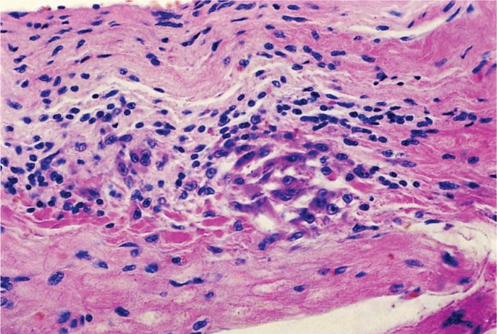
FIGURE 25–8. Aschoff nodule. Reacting lymphocytes and large mononuclear cells in myocardium demonstrate a cellular component to the immune reaction in rheumatic fever. (reproduced with permission from Connor Dh, Chandler FW, Schwartz DQ, et al: Pathology of Infectious Diseases. Stamford Ct: appleton & Lange, 1997.)
Cell-mediated immunity responses include cytotoxic lymphocytes
Genetic factors are probably also important in ARF because only a small percentage of individuals infected with GAS develop the disease. Attack rates have been highest among those of lower socioeconomic status and vary among those of different racial origins. The gene for an alloantigen found on the surface of B lymphocytes occurs four to five times more frequently in patients with rheumatic fever than in the general population. This further suggests a genetic predisposition to hyperreactivity to streptococcal products.
Alloantigens are associated with hyperreactivity to streptococci
Acute Glomerulonephritis
The renal injury of acute glomerulonephritis is caused by deposition in the glomerulus of antigen-antibody complexes with complement activation and consequent inflammation. This is a type III hypersensitivity (Chapter 2). The M proteins of some nephritogenic strains have been shown to share antigenic determinants with glomeruli, which suggests an autoimmune mechanism similar to rheumatic fever. Streptokinase has also been implicated both through molecular mimicry and through its plasminogen activation capacity.
Autoimmune reactions to M protein or streptokinase are implicated
IMMUNITY
It has long been known that an antibody directed against M protein is protective for subsequent GAS infections. This protection, however, is only for subsequent infection with strains of the same M type. This is called type-specific immunity. This protective IgG is directed against factor H-binding epitopes in the amino-terminal regions of the molecule and reverses the antiphagocytic effect of M protein. Streptococci opsonized with type-specific antibody bind complement C3b by the classical pathway, facilitating phagocyte recognition. There is evidence that mucosal IgA is also important in blocking adherence, whereas the IgG is able to protect against invasion. Unfortunately, because there are over 100 M types, repeated infections with new M types occur. Eventually, immunity to the common M types is acquired and infections become less common in adults. In ARF patients, it is the hyperreaction seen in each episode that produces the lesions associated with rheumatic heart disease.
Type-specific IgG reverses antiphagocytic effect of M protein
Repeated infections and ARF are due to many M types
 GROUP A STREPTOCOCCAL INFECTIONS: CLINICAL ASPECTS
MANIFESTATIONS
 Streptococcal Pharyngitis
Although it may occur at any age, streptococcal pharyngitis occurs most frequently between the ages of 5 and 15 years. The illness is characterized by acute sore throat, malaise, fever, and headache. Infection typically involves the tonsillar pillars, uvula, and soft palate, which become red, swollen, and covered with a yellow-white exudate. The cervical lymph nodes that drain this area may also become swollen and tender. This clinical syndrome overlaps with viral pharyngitis taking place at the same age.
Sore throat, fever, malaise
Overlaps with viral pharyngitis
GAS pharyngitis is usually self-limiting. Typically, the fever is gone by the third to fifth day, and other manifestations subside within 1 week. Occasionally, the infection spreads locally to produce peritonsillar or retropharyngeal abscesses, otitis media, suppurative cervical adenitis, and acute sinusitis. Rarely, more extensive spread occurs, producing meningitis, pneumonia, or bacteremia with metastatic infection in distant organs. In the preantibiotic era, these suppurative complications were responsible for a mortality rate of 1% to 3% after acute streptococcal pharyngitis. Such complications are much less common now, and fatal infections are rare.
Spread beyond the pharynx now uncommon
 Impetigo
The primary lesion of streptococcal impetigo is a small (up to 1 cm) vesicle surrounded by an area of erythema. The vesicle enlarges over a period of days, becomes pustular, and eventually breaks to form a yellow crust. The lesions usually appear in 2- to 5-year-old children on exposed body surfaces, typically the face and lower extremities. Multiple lesions may coalesce to form deeper ulcerated areas. Although S aureus produces a clinically distinct bullous form of impetigo, it can also cause vesicular lesions resembling streptococcal impetigo. Both pathogens are isolated from some cases.
Exposed skin of 2- to 5-year-old children
Tiny pustules may combine to form ulcers
 Erysipelas
Erysipelas is a distinct form of streptococcal infection of the skin and subcutaneous tissues, primarily affecting the dermis. It is characterized by a spreading area of erythema and edema with rapidly advancing, well-demarcated edges, pain, and systemic manifestations, including fever and lymphadenopathy. Infection usually occurs on the face (Figure 25–9), and a previous history of streptococcal sore throat is common.
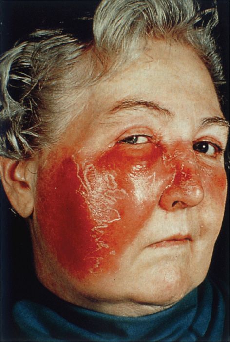
FIGURE 25–9. Streptococcal erysipelas. The diffuse erythema and swelling in the face of this woman are characteristic of GAS cellulitis at any site. (Reproduced with permission from Connor DH, Chandler FW, Schwartz DQ, et al: Pathology of Infectious Diseases. Stamford Ct: Appleton & Lange, 1997.)
Stay updated, free articles. Join our Telegram channel

Full access? Get Clinical Tree


