Patient Story
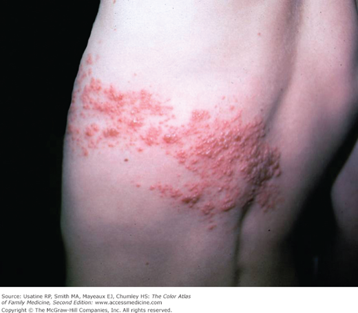
A 14-year-old boy presents with deep burning pain and a vesicular eruption in a band starting at the left chest and ending just across the midline of the back (Figure 124-1). The varicella-zoster virus (VZV) leaves the dorsal root ganglion to travel down the spinal nerves to the cutaneous nerves of the skin. The vesicles do cross the midline by a few centimeters because the posterior primary ramus of the spinal nerve includes a small cutaneous medial branch that reaches across the midline.1 The boy was treated with analgesics and an antiviral medication. The zoster healed with scarring.
Introduction
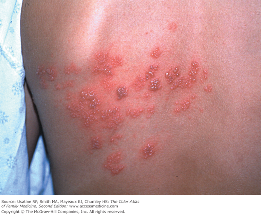
Herpes zoster (shingles) is a syndrome characterized by a painful, usually unilateral vesicular eruption that develops in a restricted dermatomal distribution (Figures 124-1 and 124-2).2,3
Epidemiology
- According to the Centers for Disease Control and Prevention (CDC), 32% of persons in the United States will experience zoster during their lifetimes accounting for about 1 million cases annually.4 Older age groups account for the highest incidence of zoster. Approximately 4% of patients will experience a second episode of herpes zoster.5
- More zoster cases have been observed among women, even when controlling for age.6
- Herpes zoster occurs more frequently and more severely in immunosuppressed patients, including transplantation patients.
Etiology and Pathophysiology
- After primary infection with either chickenpox or vaccine-type VZV, a latent infection is established in the sensory dorsal root ganglia. Reactivation of this latent VZV infection results in herpes zoster (shingles).
- Both sensory ganglia neurons and satellite cells surrounding the neurons serve as sites of VZV latent infection. During latency, the virus only expresses a small number of viral proteins.
- How the virus emerges from latency is not clearly understood. Once reactivated, virus spreads to other cells within the ganglion. The dermatomal distribution of the rash corresponds to the sensory fields of the infected neurons within the specific ganglion.3
- Loss of VZV-specific cell-mediated immune response is responsible for reactivation.3
- The pain associated with zoster infections and postherpetic neuralgia (PHN) is thought to result from injury to the peripheral nerves and altered central nervous system processing.
- The most common complications are PHN and bacterial superinfection that can delay healing and cause scarring of the zoster lesions.
- Approximately 19% of patients develop complications that may include:7
- PHN—The most common complication is seen in 10% at 90 days7 (see below).
- Ocular complications, including uveitis and keratitis (seen in 4%)7 (see Chapter 125, Zoster Ophthalmicus).
- Bell palsy and other motor nerve plastic (seen in 3%).7
- Bacterial skin infection (seen in 2%).7
- Meningitis caused by central extension of the infection.
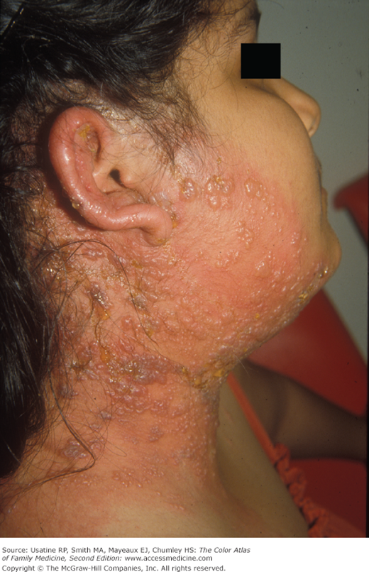
- Herpes zoster oticus (Ramsay Hunt syndrome) (Figure 124-3) includes the triad of ipsilateral facial paralysis, ear pain, and vesicles in the auditory canal and auricle.8 Disturbances in taste perception, hearing (tinnitus, hyperacusis), lacrimation, and vestibular function (vertigo) may occur.
- Other rare complications may include acute retinal necrosis, transverse myelitis, encephalitis, leukoencephalitis, contralateral thrombotic stroke syndrome, and granulomatous vasculitis.9
- PHN—The most common complication is seen in 10% at 90 days7 (see below).
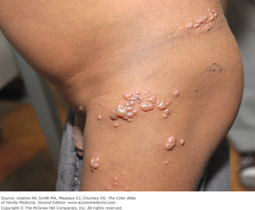
- Immunosuppressed patients are at increased risk for complications, including severe complications such as broader dermatomal involvement (Figure 124-4), disseminated infection, visceral involvement, pneumonitis, and/or meningoencephalitis.
- PHN is the persistence of pain, numbness, and/or dysesthesias precipitated by movement or in response to nonnoxious stimuli in the affected dermatome for more than 1 month after the onset of zoster. The incidence of PHN in the general population is 1.38 per 1000 person-years, and it occurs more commonly in individuals older than age 60 years and in immunosuppressed individuals.3
- In a large study, rates of zoster-associated pain (PHN) persisting at least 90 days were:
- Ten percent overall; 12% in women and 7% in men.
- Ages 22 to 59 years—Five percent overall; 6% in women and 5% in men.
- Ages 60 to 69 years—Ten percent overall; 14% in women and 5% in men.
- Ages 70 to 79 years—Seventeen percent overall; 18% in women and 15% in men.
- Age 80 years and older—Twenty percent overall; 23% in women and 13% in men.7
- Ten percent overall; 12% in women and 7% in men.
Risk Factors
- Older age.
- Underlying malignancy.
- Disorders of cell-mediated immunity.
- Chronic lung or kidney disease.
- Autoimmune disease.
- Age older than 60 years.
- Negative vaccine status.
Stay updated, free articles. Join our Telegram channel

Full access? Get Clinical Tree


