Xanthelasma Xanthelasmas (and planar xanthomas) consist of a sheet-like infiltrate of foamy macrophages involving the dermis and surrounding adnexal structures. Small areas of chronic inflammation may be present, but fibrosis and cholesterol clefts are not typical.
Eruptive Xanthoma Eruptive xanthoma is characterized by sheets of macrophages within the dermis. Extravascular lipid deposits with a blue-gray amorphous appearance are often seen between dermal collagen bundles. A sparse perivascular inflammatory infiltrate can also be seen.
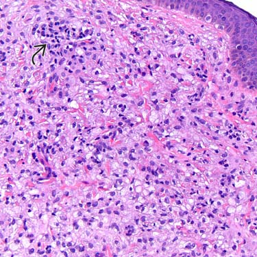
Early Eruptive Xanthoma In contrast to other forms of xanthoma, the cells in eruptive xanthoma are nonfoamy in early lesions. Note the rare foamy macrophages and extravascular lipid .
Older Eruptive Xanthoma Older eruptive xanthomas accumulate more foamy macrophages and consist of a mixture of foamy and nonfoamy cells. Again, note areas of lace-like blue-gray extravascular lipid between the dermal collagen bundles .

 may be present, but fibrosis and cholesterol clefts are not typical.
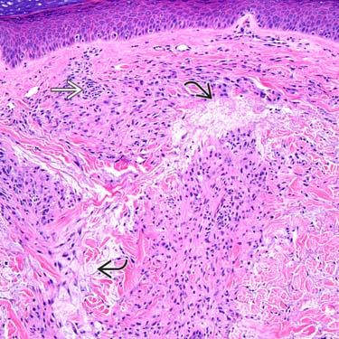
 between dermal collagen bundles. A sparse perivascular inflammatory infiltrate
  can also be seen.
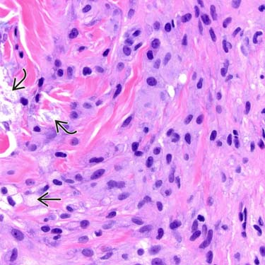
 and extravascular lipid
  .
.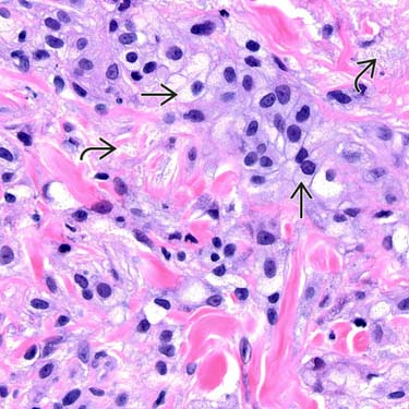
 and consist of a mixture of foamy and nonfoamy cells. Again, note areas of lace-like blue-gray extravascular lipid between the dermal collagen bundles
  .
.


