Vitamin K1


Vitamin K1


John W. Suttie


Vitamin K was discovered in 1929 by Henrik Dam (1), when he noted that chicks ingesting diets that had been extracted with nonpolar solvents to remove cholesterol developed subdural or muscular hemorrhages and that blood taken from these animals clotted slowly. Other investigators conducting studies of diet-related hemorrhage in animals (2), and by 1935, Dam (3) proposed the existence of a new fat-soluble factor, vitamin K. During the late 1930s, investigators established that menadione, 2-methyl-1,4-naphthoquinone, had vitamin K activity, and the vitamin was isolated from alfalfa as a yellow oil. This form, vitamin K1, was characterized as 2-methyl-3-phytyl-1,4-naphthoquinone (4), and it was synthesized by Doisy’s group at St. Louis University. The Doisy group also isolated a form of the vitamin from putrefied fish meal that was called vitamin K2 and contained an unsaturated polyprenyl side chain at the 3-position of the naphthoquinone ring. Early investigators recognized that the vitamin K activity of some sources of the vitamin, such as putrefied fish meal, was the result of bacterial synthesis, and they also realized that several different vitamers of the K2 series had differing chain length polyprenyl groups at the 3-position.

At the time that vitamin K was isolated and characterized, the only plasma proteins known to be involved in blood coagulation were prothrombin and fibrinogen. Dam et al (5) isolated a crude prothrombin fraction from chick plasma and demonstrated that the activity of this fraction was decreased when it was obtained from a vitamin K-deficient chick. The hemorrhagic condition resulting from obstructive jaundice or biliary problems was also shown to be caused by poor utilization of vitamin K, and these bleeding episodes were initially specifically attributed to a lack of prothrombin. A real understanding of thrombus formation and of the various soluble and cellular factors involved in regulating the generation of thrombin from prothrombin did not begin until the mid-1950s. As factors VII, IX, and X were discovered through the study of patients with clotting disorders, these factors were shown to depend on vitamin K for synthesis. For a considerable time, these three factors and prothrombin were the only proteins known to require vitamin K for their synthesis.


CHEMICAL STRUCTURE AND NOMENCLATURE

The term vitamin K is used as a generic descriptor of 2-methyl-1,4-naphthoquinone (menadione or vitamin K3) and all derivatives of this compound that exhibit an antihemorrhagic activity in animals fed a vitamin K-deficient diet (Fig. 20.1). The major dietary source of vitamin K is green plants and is generally called vitamin K1, but it is preferably called phylloquinone (USP phytonadione). The compound, 2-methyl-3-farnesylgeranyl-1,4-naphthoquinone), first isolated from putrefied fish meal, is one of a series of vitamin K compounds with unsaturated side chains called multiprenylmenaquinones that
are produced by a limited number of anaerobic bacteria and are present in large quantities in the lower bowel. This particular menaquinone (MK) has 7 isoprenoid units, or 35 carbons, in the side chain; it was once called vitamin K2, but that term is currently used to describe any of the vitamers with an unsaturated side chain, and this compound would be identified as MK-7. Vitamins of the MK series with up to 13 prenyl groups have been identified, but the predominant forms found in the gut are MK-7 through MK-9. MK-4 (2-methyl-3-geranylgeranyl-1,4-naphthoquinone) can be formed in animal tissues by alkylation of menadione (6) and is the biologically active tissue form of the vitamin used when menadione is taken as a dietary supplement.


Fig. 20.1. Structures of vitamin K active compounds. Phylloquinone (vitamin K1) synthesized in plants is the main dietary form of vitamin K. Menaquinone-9 is a prominent member of a series of menaquinones (vitamin K2) produced by intestinal bacteria, and menadione, vitamin K3, is a synthetic compound that can be converted to menaquinone-4 by animal tissues.


SOURCES AND UTILIZATION OF VITAMIN K


Analysis, Food Content, and Bioavailability

Standardized procedures suitable for the assay of the vitamin K content of foods are available, and sufficient values have been obtained (7) to provide a reasonable estimate of dietary intake of the vitamin (
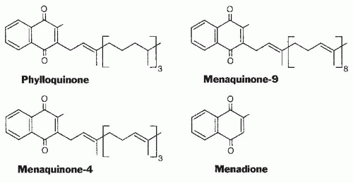
Table 20.1). Green, leafy vegetables are the foods with the highest phylloquinone content in most diets. Foods providing substantial amounts of the vitamin to most of the population are spinach (380 µg/100 g), broccoli (180 µg/100 g), and iceberg lettuce (35 µg/100 g). Fats and oils also contribute to the daily vitamin K intake of many individuals.

The phylloquinone content of oils varies considerably; soybean oil (190 µg/100 g) and canola oil (130 µg/100 g) have a high content, and corn oil (3 µg/100 g) is a poor source. The source of fat or oil has a major influence on the vitamin K content of margarine and prepared foods with a high fat content. The process of hydrogenation to convert plant oils to solid margarines or shortening converts some of the phylloquinone to 2′,3′-dihydrophylloquinone with a completely saturated side chain. The biologic activity of this form of the vitamin is lower than that of phylloquinone but has not been accurately determined. Investigators have found that the intake of this form of the vitamin by the US population is 20% to 25% that of phylloquinone (8).

The bioavailability of phylloquinone from various foods in human subjects has been difficult to assess. Initial studies compared the increase in plasma phylloquinone from the consumption of green vegetables with that of pure phylloquinone. These limited studies suggested that the bioavailability of phylloquinone from various vegetable
sources should not be considered more than 15% to 20% as available as phylloquinone consumed as a supplement. The availability of phylloquinone from vegetable oil added to corn oil was found to be about twice that from broccoli. The use of stable isotopes labeled phylloquinone should result in more accurate measurements of bioavailability (9, 10, 11). These findings demonstrate that meal composition is an important factor (12).


TABLE 20.1 PHYLLOQUINONE CONCENTRATION OF COMMON FOODSa


FOOD ITEM


µg/100 g


FOOD ITEM


µg/100 g


Vegetables


Fats and Oils


Collards


440


Soybean oil


193


Spinach


380


Canola oil


127


Salad greens


315


Cottonseed oil


60


Broccoli


180


Olive oil


55


Brussels sprouts


177


Margarine


42


Cabbage


145


Butter


7


Bib lettuce


122


Corn oil


3


Asparagus


60


Okra


40


Prepared foods


Iceberg lettuce


35


Salad dressings


100


Green beans


33


Coleslaw


80


Green peas


24


Mayonnaise


41


Cucumbers


20


Beef chow mein


31


Cauliflower


20


Muffins


25


Carrots


10


Doughnuts


10


Tomatoes


6


Potato chips


15


Potatoes


1


Apple pie


11


French fries


5


Protein sources


Macaroni/cheese


5


Dry soybeans


47


Lasagna


5


Dry lentils


22


Pizza


4


Liver


5


Hamburger/bun


4


Eggs


2


Hot dog/bun


3


Fresh meats


<1


Baked beans


3


Fresh fish


<1


Bread


3


Whole milk


<1


aMedian values.


Modified from Food and Nutrition Board, Institute of Medicine. Dietary Reference Intakes for Vitamin A, Vitamin K, Arsenic, Boron, Chromium, Copper, Iodine, Iron, Manganese, Molybdenum, Nickel, Silicon, Vanadium, and Zinc. Washington DC: National Academy Press, 2001.


A limited number of foods, mainly cheeses, do contain a significant (50 to 70 µg/100 g) amount of long-chain MKs, and a fermented soybean product, natto, that is consumed mainly in the Japanese market contains nearly 1000 µg/100 g of MK-7. Limited data indicate that the absorption of longchain MKs may be substantially higher than the absorption of phylloquinone from green vegetables (13).


Absorption and Transport of Vitamin K

Phylloquinone, the predominant dietary form of the vitamin, is absorbed from the intestine through the lymphatic system (14), and absorption is decreased in patients with biliary insufficiency or various malabsorption syndromes. Phylloquinone in plasma is predominantly carried by the triglyceride-rich lipoprotein fraction containing very-lowdensity lipoproteins and chylomicron remnants, although some is located in the low-density lipoprotein and highdensity lipoprotein fractions (15). Plasma phylloquinone concentrations in a physiologically normal population have been shown to have a mean of approximately 1.0 nmol/L (˜0.45 ng/mL), with a wide range in values from 0.3 to 2.6 nmol/L (16). As expected from this route of transport, plasma phylloquinone concentrations are strongly correlated with plasma lipid levels (17).

The major route of entry of phylloquinone into tissues appears to be through clearance of chylomicron remnants by apolipoprotein E (ApoE) receptors. The polymorphism of ApoE influences fasting plasma phylloquinone concentrations. This response is correlated with the hepatic clearance of chylomicron remnants from the circulation, with ApoE2 having the slowest rate of removal (18). The secretion of phylloquinone from the liver and the process by which the vitamin moves among organs are not yet understood.

The total human body pool of phylloquinone is very small, and turnover is rapid. A peak of circulating phylloquinone concentration following absorption has been shown to be rapidly decreased (half-life ˜15 minutes), followed by a slower decrease (half-life ˜2.5 hours) (10). Although the total amount of vitamin K is relatively high, long-chain MKs, rather than phylloquinone, are the major source of the vitamin in liver (2). Data based on liver biopsies of patients fed diets very low in vitamin K before surgery indicate that approximately two thirds of hepatic phylloquinone was lost in 3 days (19). These findings are consistent with a small pool size of phylloquinone that turns over very rapidly. The large amount of MKs in the liver, however, turns over at a much lower rate.

The major route of ingested phylloquinone excretion is through the feces, and very little unmetabolized vitamin is excreted. Many details of the metabolic transformation of the vitamin are currently lacking, but investigators have shown that the side chains of phylloquinone and MK-4 are shortened to seven or five carbon atoms yielding a carboxylic acid group at the end (14, 20). These 5C and 7C-aglycones, which are the major metabolites of phylloquinone, are excreted in the urine at concentrations that are related to the intake of the vitamin (21). Studies have also shown that glucuronides of menadione are excreted in urine at an amount that is positively related to phylloquinone (22). The mechanism by which menadione is cleaved from various sources of vitamin K or its metabolites is not known. Evidence indicates the existence of numerous other unidentified metabolites, and it also shows that treatment of patients with warfarin, which results in a substantial conversion of the body pool of phylloquinone to phylloquinone-2,3-epoxide, leads to the generation of new metabolites.


Utilization of Menaquinones from the Large Bowel

Substantial amounts of vitamin K in the form of longchain MKs are known to be present in the human gut. Relatively few of the bacteria that comprise the normal intestinal flora are major producers of MKs. Obligate anaerobes of the Bacteroides (B. fragilis), Eubacterium, Propionibacterium, and Arachnia genera are major producers, however, as are facultatively anaerobic organisms such as Escherichia coli. The amount of vitamin K in the gut can be quite large, and the amounts found in total intestinal tract contents from five patients who underwent colonoscopy ranged from 0.3 to 5.1 mg (23), with MK-9 and MK-10 the major contributors. These amounts are considerably larger than the daily dietary requirement for the vitamin, which is less than 100 µg/day. Long-chain MKs, mainly MK-6, MK-7, MK-10, and MK-11, are present at very low levels in plasma, but they have been found in human liver at levels that greatly exceed the phylloquinone concentration (24).

A major question remaining is how these very lipophilic compounds that are present as constituents of bacterial membranes are absorbed from the lower bowel. Little evidence on the route of absorption and transport of these vitamins to the liver is available.

Vitamin K deficiency in the adult human that is characterized by vitamin K-responsive hypoprothrombinemia is a very rare condition, and numerous case reports of antibiotic-induced hypoprothrombinemia are often cited as evidence of the importance of bacterial MKs. These antibiotic-induced hypoprothrombinemias have historically been presumed to result from a decrease in the synthesis of MKs by gut organisms (25), with the underlying assumption that MKs are important in satisfying at least a portion of the normal human requirement for vitamin K. In nearly all these case reports, however, evidence of
decreased MK synthesis in the presence of antibiotic treatment is lacking, and the drugs themselves may have influenced hemostatic control. The difficulty in producing a clinically significant deficiency in human subjects, such as an increased prothrombin time (PT) by dietary restriction, and the known rapid turnover of the body phylloquinone pool strongly suggest that MKs do contribute to maintaining adequate vitamin K status (24), but the magnitude of the contribution cannot be determined with the available data.


VITAMIN K-DEPENDENT PROTEINS


Hemostasis-Related Plasma Proteins

Prothrombin, the circulating zymogen of the procoagulant thrombin, was the first protein shown to depend on vitamin K for its synthesis. Prothrombin was also the first protein demonstrated to contain γ-carboxyglutamic acid (Gla) residues. Plasma clotting factors VII, IX, and X were all initially identified because their activity was decreased in the plasma of a patient with a hereditary bleeding disorder (26), and these factors were subsequently shown to depend on vitamin K1 for their synthesis. Until the mid-1970s, these four vitamin K-dependent clotting factors were the only proteins known to require this vitamin for their synthesis.

A complex series of events (Fig. 20.2), which lead to the generation of thrombin by proteolytic activation of protease zymogens (27, 28), is essential for hemostasis. The vitamin K-dependent clotting factors are involved in these activation and propagation events through membraneassociated complexes with each other and with accessory proteins. All these proteins contain a number of Gla residues, and their amino terminal Gla domain is very homologous, with 10 to 13 Gla residues in each, in essentially the same position as in prothrombin.

In addition to the classic vitamin K-dependent proteins, three more Gla-containing plasma proteins with similar homology have been discovered. Protein C and protein S participate in thrombin-initiated inactivation of factor V and therefore play an anticoagulant rather than a procoagulant role in normal hemostasis (29). In addition to the Gla domain, with approximately 40 residues, the vitamin K-dependent proteins have other common features. The Gla domain of prothrombin is followed by two kringle domains, which are also found in plasminogen, and a serine protease domain. Factors VII, IX, and X and protein C contain two epidermal growth factor domains and a serine protease domain, whereas protein S contains four epidermal growth factor domains but is not a serine protease. The function of the seventh Gla-containing plasma protein (protein Z), which is not a protease zymogen, has been shown to have an anticoagulant function under some conditions (30). Because these proteins play a critical role in hemostasis, they have been extensively studied; the cDNA and genomic organization of each of them is well documented, and many genetic variants of these proteins have been identified as risk factors in coagulation disorders (31).


Fig. 20.2. Vitamin K-dependent clotting factors involved in blood coagulation. The vitamin K-dependent procoagulants (prothrombin and factors VII, IX, and X) circulate as zymogens of serine proteases until converted to their active (subscript a) forms. Initiation of this process occurs when vascular injury exposes tissue factor to blood (extrinsic pathway). The product of the activation of one factor can activate a second zymogen, and this cascade effect results in the rapid activation of prothrombin to thrombin and the subsequent conversion of soluble fibrinogen to the insoluble fibrin clot. Some of the steps in this activation involve an active protease, a second vitamin K-dependent protein substrate, and an additional plasma protein cofactor (circles) to form a calcium (Ca2+)-mediated association with a phospholipid (PL) surface. The formation of activated factor X can also occur through thrombin activation of factor XI and subsequently factor IX (intrinsic pathway). The other two vitamin K-dependent proteins participate in hemostatic control as anticoagulants, not procoagulants. Protein C is activated by thrombin (factor IIa) in the presence of an endothelial cell protein called thrombomodulin (TM). Activated protein C functions in a complex with protein S to inactivate factors Va and VIIIa and to limit clot formation.

Jul 27, 2016 | Posted by in PUBLIC HEALTH AND EPIDEMIOLOGY | Comments Off on Vitamin K1

Full access? Get Clinical Tree

Get Clinical Tree app for offline access