Vitamin A1


Vitamin A1


A. Catharine Ross


HISTORICAL OVERVIEW

More than 2000 years ago, Egyptian and Greek physicians recognized that liver, now known as a concentrated source of vitamin A, can cure the condition known as night blindness (1). The modern era of vitamin A research began in 1913 with the independent discoveries by Osborne and Mendel and McCollum and Davis of “fat-soluble A,” later named vitamin A. These investigators observed that young rats that were fed diets containing lard or olive oil as the only fat lost weight and then died, whereas rats fed small amounts of certain “lipins,” extracted from foods such as butter, eggs, and cod liver oil, survived and began to grow again. Numerous discoveries in the next decades linked a deficiency of vitamin A to abnormalities of the eye (xerophthalmia), tissue differentiation, reproduction, and immune function. In the 1930s, vitamin A (retinol) was first synthesized de novo by Karrer, and a decade later, Arens and van Dorp synthesized the carboxylic acid form of vitamin A, retinoic acid (RA). Investigators showed that RA can substitute for retinol in many ways, but not in vision. In the 1950s and 1960s, Wald, Hubbard, and others discovered the role of vitamin A in vision and showed that “retinene” (retinal) is the essential light-absorbing component of rhodopsin (2). These basic discoveries paved the way for understanding that vitamin A is a precursor for the synthesis of two main active metabolites: 11-cis-retinal and all-trans-RA.

Research since the 1980s elucidated a mechanism of action of RA, through its binding to nuclear RA receptors (RARs), which together with retinoid X receptors (RXRs), regulate the expression of specific target genes (3, 4). Understanding the role of RA in gene expression led, in turn, to great interest in using RA and synthetic analogs (collectively termed retinoids) as therapeutic agents for the prevention or treatment of skin disorders, certain leukemias, and other diseases. In the area of public health, vitamin A supplementation programs have been shown to reduce child mortality. Vitamin A is now a key part of the global strategy to reduce child mortality as stated in United Nations Children’s Fund’s (UNICEF) Millennium Development Goals (5, 6).


TERMINOLOGY, CHEMISTRY, AND ANALYSIS

Vitamin A is a nutritional term for retinol and related compounds with its biologic activities. Dietary vitamin A can be consumed either as preformed vitamin A (retinol)
or provitamin A, namely, β-carotene and certain other carotenoids. The vitamin A molecule contains a β-ionone ring, a conjugated polyene side chain, and a terminal functional group. The parent molecule, all-trans-retinol (Fig. 17.1A), can be esterified with long-chain fatty acids to form retinyl esters (RE). Much of the world’s vitamin A is now produced commercially and is used in the production of animal feeds, nutritional supplements, and food fortification. The major synthetic forms of vitamin A are retinyl palmitate, which is identical to the major RE in most animal tissues, and retinyl acetate used in supplements.


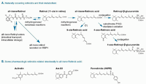
Fig. 17.1. A. Structures and metabolism of major naturally occurring retinoids. B. Pharmacologic retinoids structurally related to all-trans-retinoic acid. RBP, retinol-binding protein.

Variations of retinol having “partial” vitamin A activity also exist in nature. α-Retinol has a double bond between ring carbons 4 and 5 instead of carbons 5 and 6; it is found in tropical oils such as red palm oil and in carrots, and it has approximately one half the bioactivity of retinol (7). Vitamin A2 (chemically 3,4-didehydroretinol) is present in fresh-water fish, is also a metabolite of vitamin A in human skin, and has approximately 40% the bioactivity of retinol.

Through various steps in metabolism, discussed later, retinol is sequentially oxidized, first to retinal and then to RA. 11-cis-Retinal is the predominant form in the retina (2). All-trans-RA is the principal metabolite that regulates gene expression (3, 4), whereas 9-cis-RA and 13-cis-RA, as discussed later, are of somewhat uncertain function. Other naturally occurring retinoids contain additional substituents in the form of keto, hydroxyl, or epoxide groups. Both RA and retinol can be conjugated with glucuronic acid, which renders the molecule water soluble.

Additionally, many synthetic analogs (see Fig. 17.1B) have pharmacologic activity, such as Am80/580 (8), hydroxyphenylretinamide (fenretinide) (9), and acitretin (10). Over time, the term retinoids has come to include all of the natural vitamin A and structurally related analogs as shown in Figure 17.1.


Methods of Analysis

Most methods of retinoid analysis are based on solvent extraction to release the retinoids from proteins and other lipids in the sample, followed by chromatographic separation, generally by high-performance liquid chromatography (HPLC) with detection by ultraviolet absorption at a wavelength optimized for each compound (11, 12, 13), or, increasingly, by single or tandem mass spectrometry (LC-MSMS) (14). During storage and analysis, the sample must be protected from light and oxygen to prevent isomerization and oxidation. For quantification of total retinol in plasma and tissues, saponification is commonly used to convert the RE to retinol first, whereas without saponification, the “free” retinol and RE portions can each be determined (15).


DIETARY SOURCES AND UNITS

All vertebrates require vitamin A, but none can synthesize it de novo. The nutritional need for vitamin A can be met by either preformed vitamin A (i.e., retinol and RE present in the tissues of animals, which metabolize plantderived precursors) or provitamin A, specifically certain
carotenoids that are produced exclusively by plants, fungi, and bacteria. The highest concentrations of preformed vitamin A are found in liver and fish liver oils and other organ meats; lower levels are present in milk and eggs. Foods fortified with RE or β-carotene such as milk, margarine, and breakfast cereals are also significant sources (16). In the United States, approximately two thirds of vitamin A intake is preformed, partly from nutritional supplements. In the developing world, vitamin A is consumed mainly as provitamin A carotenoids (see the chapter on carotenoids for food sources, metabolism, and formation of vitamin A from β-carotene).


Nutritional Units

Because foods differ in their contents of preformed and provitamin A, and because the bioavailability of carotenoids differs between pure isolated compounds and food-bound compounds, the amount of vitamin A in foods must be expressed in terms of equivalents. The international unit (IU) was used for many years; by convention, 1 IU = 0.30 µg all-trans-retinol or 0.6 µg all-trans-β-carotene. The IU is still found on supplement labels, but it is outdated for calculating vitamin A in foods because it does not take into account bioavailability. In 1967, the IU was replaced by the retinol equivalent (17), which takes into account the differences in vitamin A bioactivity between β-carotene and other provitamin A carotenoids. One retinol equivalent equals 1 µg retinol, 6 µg β-carotene, and 12 µg of other provitamin A carotenoids. In 2001, after studies had shown that the bioavailability of carotenoids in foods is lower than previously thought, a new unit, the retinol activity equivalent (RAE), was adopted (18): 1 µg RAE equals 1 µg of pure all-trans-retinol, 2 µg of pure all-trans-β-carotene in oil (a highly absorbable form), 12 µg of food-based all-trans-β-carotene in foods (from which absorption is less), and 24 µg of other, food-based, all-trans-provitamin A carotenoids (18). Nutritionally, one molecule of retinol and one molecule of any form of RE are equivalent.


Dietary Reference Intakes

Dietary reference intake (DRI) values are expressed in micrograms RAE/day (Table 17.1). The estimated average requirement (EAR) and recommended dietary allowance (RDA) values for vitamin A for the United States and Canada were established by the Institute of Medicine (IOM) in 2001. The EAR, which is defined as the middle of the requirement distribution for the population and which serves as a basis for the RDA, was determined using a factorial method that takes into account the proportion of vitamin A stores that are lost per day (0.5%), an estimation of minimum acceptable liver vitamin A reserves (20 µg/g) (19), and the efficiency of storage of ingested vitamin A (18). The EAR for pregnant women was calculated by including an additional amount to cover the transfer of vitamin A to the growing fetus, whereas for lactation, an additional amount was added to cover the secretion of vitamin A in breast milk (18). For all age-sex groups, RDA values were calculated by adding an assumed coefficient of variation of 20% to the EARs. For infants 0 to 6 and 7 to 12 months of age, however, the recommended intake is expressed as an adequate intake (AI) based on multiplying average values for the concentration of vitamin A in breast milk by the average milk volume for each of the two age groups.


TABLE 17.1 DIETARY REFERENCE INTAKE VALUES FOR VITAMIN A BY LIFE STAGE GROUP


LIFE STAGE GROUP


RDA (µg RAE/d)a


AIb (µg/d)


ULc (µg PREFORMED RETINOL/d)


MALE


FEMALE


0-6 mo


400


600


7-12 mo


500


600


1-3 y


300


300


600


4-8 y


400


400


900


9-13 y


600


600


1,700


14-18 y


900


700


2,800


>18 y


900


700


2,800/3,000d


Pregnancy


14-18 y


750


2,800


19-50 y


770


3,000


Lactation


14-18 y


1,200


2,800


19-50 y


1,300


3,000


a RDA, recommended dietary allowance, is the intake that meets the nutrient need of almost all (97% to 98%) individuals in a group. RAE, retinol activity equivalent.

b AI, adequate intake, is the observed average or experimentally determined intake by a defined population or subgroup that appears to sustain a defined nutritional status, such as growth rate, normal circulating nutrient values, or other functional indicators of health. The AI is used if sufficient scientific evidence is not available to derive an estimated average requirement (EAR). For healthy infants receiving human milk, the AI is the mean intake. The AI is not equivalent to an RDA.

c UL, tolerable upper intake limit, is the highest level of daily nutrient intake that is likely to pose no risk of adverse health effects for almost all individuals in the specified life stage group.

d UL equals 2800 µg/day for women and 3000 µg/day for men more than 18 years old.


Reprinted with permission from Food and Nutrition Board, Institute of Medicine. Dietary Reference Intakes for Vitamin A, Vitamin K, Arsenic, Boron, Chromium, Copper, Iodine, Iron, Manganese, Molybdenum, Nickel, Silicon, Vanadium, and Zinc. Washington, DC: National Academy Press, 2001.


Reference intakes have also been established by other nations and by the Food and Agriculture Organization/World Health Organization (FAO/WHO); these values tend to be lower than the RDAs (20). In practice, a person can obtain sufficient vitamin A from any mixture of preformed vitamin A and provitamin A carotenoids that provides the recommended level in terms of RAE/day (18).


Tolerable Upper Intake Levels

In calculating the tolerable upper intake levels (ULs), the highest level that poses no risk over long periods, the IOM committee considered only preformed vitamin A because β-carotene has not been shown to be associated with
increased risk (16, 18). The critical indicators used to specify the UL were birth defects in women of reproductive age and liver abnormalities for all other age-sex groups. The UL is specified as 3000 µg of retinol/day for both women and men, and 600, 900, 1700, and 2800 µg retinol/day for the age ranges 0 to 3, 4 to 8, 9 to 13, and 14 to 18 years, respectively (18).


METABOLISM

A schematic of whole body vitamin A metabolism is depicted in Figure 17.2. The metabolism of retinoids is directed in part by their binding to specific proteins, as discussed next, and by various enzymes that convert retinol to its storage form, mobilize REs, and oxidize retinol and retinal to RA, as subsequently discussed. Several processes in vitamin A metabolism are regulated by vitamin A metabolites, resulting in a certain level of autoregulation.


Chaperone Proteins That Facilitate Vitamin A Metabolism

Chaperones belonging to the families of retinoid-binding proteins and retinoid receptors are critical for the normal metabolism of vitamin A. Retinoid-binding proteins confer aqueous solubility to lipophilic molecules and serve as guides for the transport and metabolism of specific retinoids, whereas the nuclear receptors mediate the functions of RA.


Plasma Transport Proteins: Retinol-Binding Protein and Transthyretin

Approximately 95% of the vitamin A present in plasma is in the form of all-trans-retinol, and nearly all of it is bound to retinol-binding protein (RBP), which is sometimes referred to by its gene designation RBP4. RBP is described as holo-RBP when retinol is bound to it and as apo-RBP in the absence of retinol. RBP is a 21-kDa protein belonging to the lipocalin family that has the overall structure of a “β-barrel.” Each protein molecule binds one molecule of retinol within a hydrophobic cavity, with the hydroxyl group of retinol oriented toward the surface of RBP (21). RBP circulates bound to the protein transthyretin (TTR, previously known as prealbumin), which is one of the plasma transport proteins for thyroxine. The association between a molecule of RBP and a tetramer of TTR is noncovalent; this association serves to stabilize holo-RBP, as shown in vitro and in vivo (22, 23, 24). Some other retinoids bind to RBP but in a less stable manner. For example, the retinol analog 4-HPR binds to RBP, but the complex interacts relatively weakly with TTR. As a result, RBP is more readily lost in urine, and plasma retinol concentrations are reduced (23, 25). α-Retinol, apparently because of its more planar overall structure than retinol (formally β-retinol), does not bind effectively to RBP (26).

Although the rate of synthesis of RBP is normally high, the plasma concentration is relatively low, approximately 1 to 3 µM, at least as compared with other plasma proteins (27). This is related to the rapid turnover of RBP, with a half-life of approximately 0.5 days for the holoprotein and 4 hours for apo-RBP (24). Because the formation of the holo-RBP-TTR complex (23) increases its molecular weight, to approximately 75 kDa, complex formation slows the rate of loss of RBP in the kidneys.

The gene RBP4 encoding RBP covers approximately 1000 base pair of cDNA, and its mRNA is one of the most highly expressed in the liver (27), with mRNA expression localized to hepatocytes (28). RBP4 mRNA is also expressed in adipose tissue and kidney at approximately 3% to 10% of the level present in liver (27), a finding suggesting that these organs may also synthesize
RBP protein. Adipose-derived RBP may function as an adipokine and play a role in glucose homeostasis (29), and numerous studies have correlated its levels with various metabolic parameters. Whether it is a causative factor or a correlative biomarker is still unclear, however.


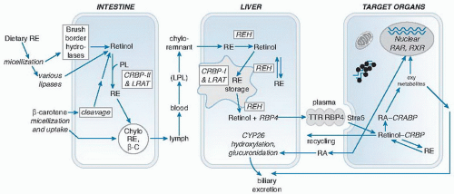
Fig. 17.2. Major reactions in retinoid metabolism. CRABP, cellular retinoic acid-binding protein; CRBP, cellular retinol-binding protein; LPL, lipoprotein lipase; LRAT, lecithin: retinol acyltransferase; PL, phospholipid; RA, retinoic acid; RAR, retinoic acid receptor; RBP, retinol-binding protein; RE, retinyl ester; REH, retinyl ester hydrolase; RXR, retinoid X receptor; Stra6, stimulated by RA gene 6; TTR, transthyretin.

The liver is the main, but not the only, site of synthesis of TTR (22). The molar concentration of TTR in plasma is higher than that of RBP, and thus most TTR circulates as the free tetramer. Numerous TTR polymorphisms are known, and although some of them affect thyroxine or RBP binding, most are associated with familial amyloidotic polyneuropathies (22).


Cellular Retinoid-Binding Proteins

Several cellular retinoid-binding proteins act as intracellular chaperones for retinol, retinal or RA (30, 31). The cellular RBPs (CRBPs) CRBP-I, II, and III belong to the fatty acid binding protein/CRBP family, are of similar size, approximately 14.6 kDa, and have a β-clam structure with a hydrophobic binding site that binds a single molecule of retinol with its hydroxyl group inward (30). CRBP-I, the most abundant form expressed in liver, kidney, testes, and other tissues, binds all-trans-retinol, whereas CRBP-II binds both all-trans-retinol and retinal and is abundant in enterocytes (32). Neither appreciably binds 9-cis-retinoids. CRBP-III and CRBP-IV are present in heart, skeletal muscle, kidney, and some other tissues, but they are less well studied (31).

The cellular RA-binding proteins (CRABP) CRABP-I and CRABP-II, which are structurally similar to the CRBP proteins, bind all-trans-RA (33). They are also expressed in tissue-specific patterns, generally at lower concentrations than CRBPs (31). Both are expressed in the developing embryo but usually not in the same cells, a finding suggesting that they perform different functions.

Two other cellular retinoid-binding proteins, cellular retinal-binding protein (CRALBP) and interstitial retinoid-binding protein (IRBP) are expressed almost exclusively in the eye (see the later section on ocular retinoid metabolism).


Nuclear Retinoid Receptors

Nuclear retinoid receptors of the RAR and RXR gene families are members of the superfamily of steroid/thyroid hormone receptors (3, 4). Each consists of three genes: RAR-α, β, and γ, and RXR-α, β, and γ, with considerable structural similarity, especially within the ligand-binding domain of each subgroup (34, 35). Tissue expression, however, differs for each receptor. The RARs bind all-trans-RA exclusively. The RARs can bind 9-cis-RA, but, alternatively, other physiologic ligands have been suggested, including unsaturated fatty acids and phytanic acid (36). Synthetic “rexinoids” selectively activate the RXRs (37). Additionally, they may also function in a ligand-independent manner (38). Functionally, the RXR and RAR bind to each other as heterodimers, which, in turn, bind to specific DNA sequences in retinoid-responsive genes, as described later. The binding of ligand, for example, of all-trans-RA to RAR, induces a conformational change in the receptor that facilitates its interaction other proteins, including coactivator or corepressor proteins, enzymes that modify chromatin-bound histones, basal transcription factors, and RNA polymerases (3, 35). The amount of retinoid receptor protein available for ligand binding can be regulated by transcription, posttranscriptional modification, proteolysis, and protein trafficking (35). The RXRs also form heterodimers with other nuclear receptors, including the vitamin D receptor, peroxisome proliferator-activated receptor (PPAR), farnesoid X receptor (FXR), liver X receptor (LXR), and receptors for certain drugs and xenobiotics (34), thus participating in various regulatory networks.

The retinoid-response elements to which RAR-RXR heterodimers bind are typically a direct repeat (DR) of the hexanucleotide sequence (A/G)/(G/T)GTCA, with either five or two intervening nucleotides, referred to as a DR-5 or DR-2, respectively, which are most often located in the 5′-regulatory region of retinoid-responsive genes. Some, however, lie in introns or outside of genes (39). CRABP-II, RAR-β, and CYP26A1 (discussed in the section on metabolism) contain one or more retinoic acid response element (RARE), which provides a means for RA to self-regulate certain aspects of its own metabolism and functions. For many genes, despite evidence of their physiologic regulation by RA, no RARE has been identified, and they could be regulated indirectly (39).


Intestinal and Hepatic Retinoid Metabolism

Metabolism is characterized by extensive interorgan trafficking of retinol, cycles of retinol esterification to form RE and of the hydrolysis of RE to regenerate retinol, and stepwise oxidative metabolism. Approximately 70% of dietary vitamin A is absorbed, even when intake is high, and this has implications for vitamin A overload and toxicity. In contrast, plasma retinol is maintained at a nearly constant level, except in states of vitamin A deficiency and excess (19). Thus, tissues are normally exposed to a wellregulated supply of plasma retinol.


Intestinal Retinol Absorption

Vitamin A absorption comprises the processes of digestion, emulsification, uptake, intracellular metabolism, and export from the intestine into the lymphatic system or portal blood (40). REs in foods must be liberated from chyme by digestive enzymes, and REs, regardless of source, must be emulsified with fatty acids and bile salts and incorporated into lipid micelles before hydrolysis by RE hydrolases (REH) and the uptake of retinol into the duodenal and jejunal enterocytes. REHs include colipasedependent pancreatic lipase (41), as well as microvillus membrane-associated enzymes. Conditions that interfere with digestion and lipid emulsification, including dietary fat less than approximately 5%, may reduce the efficiency of vitamin A absorption (18).


After uptake of free retinol by enterocytes (42), approximately 95% is esterified as REs (43). Retinol for esterification is carried by CRBP-II to the membrane-bound enzyme lecithin: retinol acyltransferase (LRAT), which transfers the fatty acid in the stereospecific nomenclature (sn)-1 position of membrane-associated phosphatidyl choline (lecithin) to retinol, thus forming RE. The composition of sn-1 fatty acids in lecithin dictates that LRAT will form a mixture of retinyl palmitate in quantities greater than stearate, oleate, and linoleate in most tissues. LRAT is an obligatory enzyme, as shown by studies in mice that lack LRAT and accumulate little RE in their tissues (44). The newly formed REs, along with triglycerides and cholesteryl esters, are packaged into the lipid core of the nascent chylomicron (41). The quantity of RE formed in the enterocyte and the amount of RE per chylomicron particle vary in direct proportion to the amount of vitamin A being absorbed and esterified at the time (45), which can vary from nil after a meal free of vitamin A up to several milligrams per gram of lipid after ingestion of a high-vitamin A meal or vitamin A supplement (45).

The trafficking of chylomicron RE is mainly determined by the metabolism of the chylomicron itself. Chylomicrons enter the lymphatic system and then the venous circulation, and they peak in concentration in plasma approximately 2 to 6 hours after meals. Whereas chylomicron triglycerides are rapidly metabolized in tissues containing lipoprotein lipase (LPL), the chylomicron remnant contains nearly all the original RE, except for a small fraction that may transfer into tissues during the LPL reaction or exchange with plasma lipoproteins. Because of the very rapid hepatic uptake of chylomicron remnants, dietary REs have a short half-life, less than 20 minutes, in the plasma of normal subjects (46). If chylomicron clearance is impaired or the absorption of RE is extremely high, then REs may be found in plasma at more than a few percent of total retinol concentration. Some tissue uptake of RE may take place through lipoprotein receptors (47), or during lipolysis by LPL (48, 49). Approximately 60% to 80% of dietary RE is taken up by the liver during the process of chylomicron remnant clearance, however.

Chylomicrons also contain a minor portion of unesterified retinol (5% to 10% of total vitamin A), which may more readily exchange with tissues and lipoproteins. Additionally, some small fraction of newly absorbed vitamin A is oxidized to polar retinoids in the intestinal mucosal cells. RA is absorbed bound to albumin (27). Portal venous blood RA increases after a dose of β-carotene (50), and most likely after vitamin A.

In isotope kinetic studies, approximately 70% to 90% of a physiologic dose of vitamin A was absorbed (51). The process of retinol absorption is relatively unregulated, and absorption is high even when the dose is very large (18), a situation that may contribute to the development of hypervitaminosis A (see later section). Chylomicrons in humans contain a minor proportion of intact β-carotene (52), but mostly RE. In rodents, nearly all carotene is cleaved and absorbed as RE.


Hepatic Metabolism

The liver plays a central role in whole body retinoid homeostasis. The RE molecules in chylomicron remnants are hydrolyzed soon after uptake by the liver (53, 54). Whereas this process appears insensitive to vitamin A status, what happens after this initial hydrolysis depends greatly on vitamin A status. In a study that traced the metabolism of chylomicron 3H-RE, in vitamin A-adequate rats most of the 3H was initially taken up by hepatocytes, but it was then transferred within 2 hours into hepatic stellate cells (HSCs) (54), which contain the CRBP-I and lecithin retinol acyltransferase (LRAT) necessary to synthesize RE and to store REs within their cytoplasmic lipid droplets (55). These REs make up approximately 50% to 85% of total body vitamin A, more than 90% as RE, in the well-nourished state (56). In contrast, in vitamin A-deficient rats, very little retinol was transferred to HSCs (54); instead, retinol rapidly appeared in the plasma compartment. That liver LRAT expression and activity decline progressively as vitamin A deficiency develops is known (45). Therefore, the reduction in hepatic LRAT is likely part of a regulatory mechanism that spares the little remaining retinol for other uses, such as secretion as holo-RBP or conversion to RA.

The proportion of CRBP as apo-CRBP also increases as vitamin A status declines, and apo-CRBP stimulates the hydrolysis of RE by REH (57). The result is that essentially all vitamin A in the liver can be mobilized and used. Conversely, when vitamin A is administered to deficient animals, holo-RBP is very rapidly secreted, and then hepatic LRAT expression increases (58), resulting in restoration of normal plasma retinol and the appearance of stored REs within a few hours. Although most studies have been conducted in mice and rats, human retinol metabolism is likely to be similar, based on a similar range of vitamin A levels in human liver specimens (59), as well as observations of a similar rapid rise in the plasma of vitamin A-deficient persons after vitamin supplementation.


Hepatic Synthesis and Secretion of Retinol-Binding Protein

Hepatocytes synthesize RBP as a 24-kDa preprotein that is cleaved during translation to form the mature 21-kDa protein (27). The movement of RBP through the secretory pathway depends on its combining with retinol to form holo-RBP (60). In vitamin A deficiency, RBP mRNA stays relatively constant, but RBP protein accumulates within the hepatocytes as apo-RBP, to be released as holo-RBP on vitamin A repletion. In vitamin A-deficient rats, the concentrations of plasma retinol and RBP rose from nearly undetectable to a level higher than normal in approximately 5 hours after vitamin A repletion and then stabilized at a normal level (61). These findings provide
the rationale for the relative dose response (RDR) test, described later, which is used clinically.


Retinoids in Plasma


Retinol

In healthy humans in the fasting state, plasma vitamin A is mainly in the form of retinol (>95%). Plasma REs are elevated transiently after vitamin A-containing meals as a result of the REs in chylomicrons and their remnants. If RE constitutes 5% to 10% of the total retinol in fasting plasma, however, then this suggests an abnormal situation, such as impaired chylomicron clearance or an excessive intake of dietary vitamin A (hypervitaminosis A; see later). Considerable variation exists among animal species in retinol transport in plasma: whereas most rodents used in laboratory research resemble humans and transport mostly of their plasma vitamin A as retinol, most of the plasma vitamin A in great apes and several species of carnivores is found as RE bound to lipoproteins (62). In dogs, plasma REs were present in the fasted state, and, surprisingly, even after weeks on a vitamin A-deficient diet (63).

Plasma retinol concentrations in adults normally range from approximately 1 to 3 µmol/L (equivalent to 28 to 86 µg/dL). Day-to-day variation is low. The molar ratio of plasma retinol to RBP is approximately 0.82, indicating that some apo-RBP is normally present in plasma (64). The median plasma retinol concentration measured in the National Health and Nutrition Examination Survey (NHANES) is age related, lower in young children than in adolescents and higher in adult men than in premenopausal women (65). After 50 years of age, values are similar in men and women. In women using oral contraceptives, the retinol concentration is 15% to 35% higher. In newborns, values are lower for premature infants than for full-term infants (66).

Plasma retinol values of less than 0.35, less than 0.70, and less than 1.05 µmol/L are often interpreted as indicating states of severe deficiency, marginal deficiency, and subclinical low vitamin A status, respectively. Based on analysis of serum retinol in NHANES from 1988 to 1994, the prevalence of low serum retinol lower than 0.70 µmol/L in all strata of the US population was very low (67). The prevalence of serum retinol lower than 1.05 µmol/L was 16.7% to 33.9% in children aged 4 to 8 years and 3.6% to 14.2% in children aged 9 to 13 years, depending on sex and racial or ethnic group, but it was higher in non-Hispanic black and Mexican American children than in non-Hispanic white children even after controlling for covariates. As reviewed in the literature (68), the WHO and other organizations use plasma retinol as one criterion for determining whether low vitamin A status is a public health problem in regions or countries.

Only gold members can continue reading. Log In or Register to continue

Stay updated, free articles. Join our Telegram channel

Jul 27, 2016 | Posted by in PUBLIC HEALTH AND EPIDEMIOLOGY | Comments Off on Vitamin A1

Full access? Get Clinical Tree

Get Clinical Tree app for offline access