• Vessel may collapse after biopsy and not appear ectatic
• Vessel may be thrombosed
• Background of solar elastosis
Top Differential Diagnoses
• Hemangioma
Multiple not single vessels
• Pyogenic granuloma
Lobular proliferation of capillaries (type of lobular capillary hemangioma)
Often overlying epidermal ulceration and serum crust with acute inflammation
• Angiokeratoma
Vessels herniate into overlying epidermis
• Lymphangioma
Ectatic lymphatic channels
Have lymphatic valves
• Angiosarcoma
• Kaposi sarcoma
Clinical Image of Venous Lake Venous lake presents as a dark blue papule on the sun-damaged skin of elderly patients. It is especially common on the lip.
Low Magnification of Venous Lake Venous lake is characterized by a single large, ectatic vessel in actinically damaged skin.
High Magnification of Venous Lake The vessel of a venous lake is lined by flattened endothelial cells that lack atypia.
Venous Lake With Thrombus This is an example of a venous lake presenting on the lip (note the squamous mucosa overlying the lesion), one of the most common locations. The vessel has a partial thrombus filling its lumen, consistent with Masson change.
TERMINOLOGY
Synonyms
• Capillary aneurysm
Definitions
• Type of telangiectasia characterized by ectatic superficial blood vessel in superficial dermis
Only gold members can continue reading. Log In or Register to continue


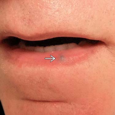
 on the sun-damaged skin of elderly patients. It is especially common on the lip.
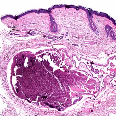
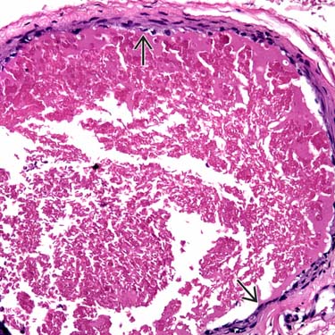
 that lack atypia.
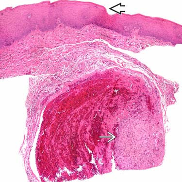
 overlying the lesion), one of the most common locations. The vessel has a partial thrombus
  filling its lumen, consistent with Masson change.


