• Clavicle: subcutaneous; palpable throughout its length • Acromion: easily palpable, traced medially to acromioclavicular joint (see Section 6.2, Shoulder and Axilla) • Coracoid process of scapula: palpable deep and lateral to deltopectoral triangle • Head of humerus: palpable deep to deltoid inferior to lateral edge of acromion when arm is rotated • Elbow: palpable medial and lateral epicondyles of humerus and visible olecranon process of ulna (see Section 6.4, Elbow and Forearm) • Head of radius: palpable as it rotates during pronation and supination on posterolateral aspect of extended elbow, just distal to lateral epicondyle • Ulnar head: visible on medial side of dorsal aspect of wrist (see Section 6.5, Wrist and Hand) • Radial styloid: palpable in anatomical snuff box on lateral side of wrist • Ulnar styloid: just distal to ulnar head with hand supinated • Pisiform: hard, round structure on anteromedial aspect of wrist • Tubercles of scaphoid and trapezium: palpable at proximal end of thenar eminence • Deltoid muscle: Overlies shoulder, giving it rounded appearance • Biceps: Bulge on anterior aspect of arm • Biceps brachii tendon: palpable in cubital fossa, lateral to midline with arm flexed • Flexor tendons: wrist and finger flexors visible distally on ventral aspect of forearm • Extensor tendons: wrist and finger flexors visible on the dorsum of hand • Thenar eminence: muscles at base of thumb (see Section 6.5, Wrist and Hand) • Hypothenar eminence: muscles at base of little finger (see Section 6.5, Wrist and Hand) • Radial artery: pulse can be felt by compressing artery against distal end of radius • Median cubital vein (see Section 6.4, Elbow and Forearm) • Cephalic vein ascends along lateral forearm and arm (see Section 6.6, Neurovasculature) • Basilic vein ascends along medial forearm and distal arm (see Section 6.6, Neurovasculature) • Clavicle: sternal and acromial ends • Sternal end articulates with manubrium of sternum • Acromial end articulates with acromion • Deltoid tubercle for attachment of deltoid muscle • Conoid tubercle for attachment of conoid ligament • Subclavian groove for attachment of subclavius muscle • Serves as a strut suspending scapula and limb with maximum freedom • Scapula: lying against posterolateral thorax • Lies posterolateral on 2nd through 7th ribs • Concave costal surface = subscapular fossa • Posterior surface divided by spine = transverse ridge of bone • Acromion = flattened lateral end of spine • Coracoid process = anterior projection above glenoid cavity • Glenoid cavity = socket for head of humerus • Suprascapular notch (scapular notch) = notch found on superior border, two thirds of way along laterally • Divided into two compartments by an articular disc • Articulation between concave facet of manubrium and concave facet of clavicle • Strengthened by anterior and posterior sternoclavicular, costoclavicular, and interclavicular ligaments • Blood supply: branches of suprascapular and internal thoracic arteries • Nerve supply: branches of supraclavicular nerve and nerve to subclavius • No demonstrable movement; muscles moving scapula cause acromion to move on clavicle • Articulation between concave facet of acromion and convex facet of clavicle • Strengthened by acromioclavicular and coracoclavicular (conoid and trapezoid) ligaments • Unites coracoid process and clavicle • Two component ligaments: conoid: vertical, in shape of inverted pyramid; trapezoid: horizontal, extends laterally to inferior surface of clavicle • Blood supply: branches of suprascapular and thoraco-acromial arteries • Nerve supply: branches of supraclavicular, lateral pectoral, and axillary nerves • Shoulder (glenohumeral) joint • Multiaxial, synovial ball-and-socket joint • Articulation of head of humerus with shallow glenoid cavity of scapula • Joint socket deepened by glenoid labrum (fibrocartilaginous ring) and supported by the rotator cuff muscles (see below) • Loose fibrous capsule encloses and contains two apertures • Between tubercles of humerus for passage of long head of biceps brachii, which attaches to supraglenoid tubercle within joint • Anterior opening, inferior to coracoid process, for communication between subscapular bursa and synovial cavity of joint • Blood supply: branches of anterior and posterior circumflex humeral arteries from axillary and suprascapular artery from subclavian • Nerve supply: branches of suprascapular, axillary, and lateral pectoral nerves • Ligaments of glenohumeral joint • Glenohumeral ligaments—strengthen capsule anteriorly • Coracohumeral ligament—strengthens joint superiorly • Transverse humeral ligament—bridges gap between greater and lesser tubercle and holds tendon of biceps brachii in place • Coraco-acromial ligament—from acromion to coracoid process, prevents displacement of humeral head superiorly • Superficial extrinsic (join axial skeleton to appendicular skeleton) • Deltoid: gives shoulder its rounded appearance, abducts arm past 15 degrees • Teres major: adducts and medially rotates arm • Teres minor: hidden by deltoid, assists lateral rotation of arm and adduction • Supraspinatus: initiates arm abduction • Infraspinatus: laterally rotates arm • Subscapularis: primary medial rotator of the arm, also adducts • Pyramid-shaped area inferior to glenohumeral joint containing important neurovascular structures to upper limb • Base: skin of armpit and axillary fascia from arm to thoracic wall • Apex: bounded by 1st rib, clavicle, and superior border of scapula • Anterior wall (anterior axillary fold): pectoralis major and minor • Posterior wall (posterior fold): subscapularis, teres major, latissimus dorsi • Medial wall: 1st through 4th ribs, serratus anterior, and intercostal muscles • Contents (see Section 6.6, Neurovasculature, for details) • Axillary artery and branches • Axillary vein and tributaries • Axillary lymph nodes (five major collections) • Invests subclavius and pectoralis minor • Continues superiorly as costocoracoid membrane (pierced by lateral pectoral nerve) • Axillary sheath: invests axillary artery and brachial plexus • Common, especially in children • Usually results from a fall on outstretched hand or direct trauma to the shoulder • Fractures of middle third are most frequently seen. • Sternocleidomastoid muscle pulls the proximal fragment superiorly and the shoulder pulls the distal fragment inferiorly. • Inflammation and calcification of the subacromial bursa resulting in pain, tenderness, and limitation of movement of the shoulder joint • Calcium deposits frequently also seen in the supraspinatus tendon. • Pain is especially severe with the arm abducted between 50 to 130 degrees (the painful arc) as the supraspinatus tendon is in contact with the inferior surface of the acromion here. • High mobility and instability of the glenohumeral joint leads to frequent dislocation. • 95% of dislocations are in anteroinferior direction, caused by excessive extension and lateral rotation of humerus (e.g., in the throwing motion). • Humeral head places stress on joint capsule, which may be torn anteriorly, with elements of the rotator cuff. • Axillary and musculocutaneous nerves may also be injured. • Posterior dislocation is uncommon but may occur during epileptic seizure or electrocution. • Musculotendinous rotator cuff may be damaged by trauma or degenerative disease. • One or more of tendons may be torn when the arm is forcefully abducted, leading to pain in the anterosuperior aspect of the shoulder. • Supraspinatus tendon is most commonly involved in degenerative tendonitis. • Leads patient’s arm to drop suddenly at approximately 90-degree abduction, when instructed to lower it slowly from a fully abducted position • Anatomical neck—circumscribes head above tubercles • Surgical neck: below tubercles—common site of fracture • Greater and lesser tubercles • Deltoid tuberosity for attachment of deltoid muscle • Radial groove on posterior surface where radial nerve and deep brachial artery traverse • Medial and lateral supracondylar ridges—widening of humerus distally as sharp ridges on either side • Prominent medial extension at distal end • Common origin of forearm extensors; radial nerve posterior • Condyle—distal end of humerus • Coronoid fossa (see also Section 6.4, Elbow and Forearm) • Medial and lateral intermuscular septa • Extend from deep surface of brachial fascia to humerus • Divide arm into anterior (flexor) and posterior (extensor) compartments • Medial septum: medial lip of intertubercular sulcus (superiorly) → medial epicondyle • Lateral septum: lateral lip of intertubercular sulcus (superiorly) → lateral epicondyle • Anterior (flexor) compartment • Continues distally as bicipital aponeurosis: triangular membrane from biceps tendon across cubital fossa and blends with antebrachial fascia over flexor muscles of forearm • Brachialis: main flexor of forearm • Posterior (extensor) compartment
Upper Limb Study Guide
6.1 Topographic Anatomy
Guide
Bones
Muscles and Tendons
Arteries and Veins
6.2 Shoulder and Axilla
Guide
Bones
Joints
Ligament
Attachments
Comment
Joint capsule
Margin of glenoid cavity → anatomical neck of humerus
Loose fibrous capsule
Weakest inferiorly
Glenohumeral
Supraglenoid tubercle → blend with fibrous capsule (superior, middle and inferior bands)
Reinforce anterior capsule
Coracohumeral
Coracoid process → greater tubercle of humerus
Strong
Transverse humeral
Bridges intertubercular groove between greater and lesser tubercles
Holds tendon of biceps brachii in intertubercular groove
Coraco-acromial
Coracoid process → acromion
Completes coraco-acromial arch protecting humeral head
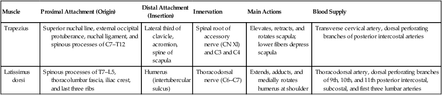
Muscles of the Scapula
Muscle
Proximal Attachment (Origin)
Distal Attachment (Insertion)
Innervation
Main Actions
Blood Supply
Trapezius
Superior nuchal line, external occipital protuberance, nuchal ligament, and spinous processes of C7–T12
Lateral third of clavicle, acromion, spine of scapula
Spinal root of accessory nerve (CN XI) and C3 and C4
Elevates, retracts, and rotates scapula; lower fibers depress scapula
Transverse cervical artery, dorsal perforating branches of posterior intercostal arteries
Latissimus dorsi
Spinous processes of T7–L5, thoracolumbar fascia, iliac crest, and last three ribs
Humerus (intertubercular sulcus)
Thoracodorsal nerve (C6–C7)
Extends, adducts, and medially rotates humerus at shoulder
Thoracodorsal artery, dorsal perforating branches of 9th, 10th, and 11th posterior intercostal, subcostal, and first three lumbar arteries
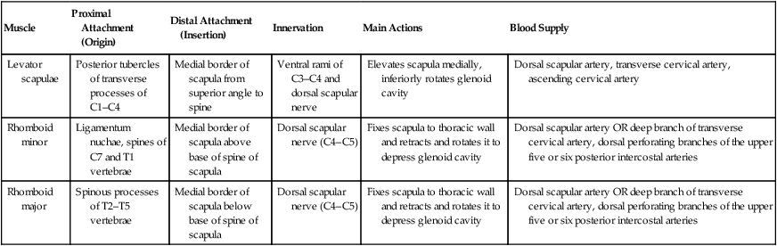
Muscle
Proximal Attachment (Origin)
Distal Attachment (Insertion)
Innervation
Main Actions
Blood Supply
Levator scapulae
Posterior tubercles of transverse processes of C1–C4
Medial border of scapula from superior angle to spine
Ventral rami of C3–C4 and dorsal scapular nerve
Elevates scapula medially, inferiorly rotates glenoid cavity
Dorsal scapular artery, transverse cervical artery, ascending cervical artery
Rhomboid minor
Ligamentum nuchae, spines of C7 and T1 vertebrae
Medial border of scapula above base of spine of scapula
Dorsal scapular nerve (C4–C5)
Fixes scapula to thoracic wall and retracts and rotates it to depress glenoid cavity
Dorsal scapular artery OR deep branch of transverse cervical artery, dorsal perforating branches of the upper five or six posterior intercostal arteries
Rhomboid major
Spinous processes of T2–T5 vertebrae
Medial border of scapula below base of spine of scapula
Dorsal scapular nerve (C4–C5)
Fixes scapula to thoracic wall and retracts and rotates it to depress glenoid cavity
Dorsal scapular artery OR deep branch of transverse cervical artery, dorsal perforating branches of the upper five or six posterior intercostal arteries
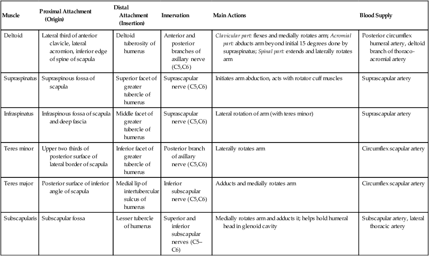
Muscle
Proximal Attachment (Origin)
Distal Attachment (Insertion)
Innervation
Main Actions
Blood Supply
Deltoid
Lateral third of anterior clavicle, lateral acromion, inferior edge of spine of scapula
Deltoid tuberosity of humerus
Anterior and posterior branches of axillary nerve (C5,C6)
Clavicular part: flexes and medially rotates arm; Acromial part: abducts arm beyond initial 15 degrees done by supraspinatus; Spinal part: extends and laterally rotates arm
Posterior circumflex humeral artery, deltoid branch of thoraco-acromial artery
Supraspinatus
Supraspinous fossa of scapula
Superior facet of greater tubercle of humerus
Suprascapular nerve (C5,C6)
Initiates arm abduction, acts with rotator cuff muscles
Suprascapular artery
Infraspinatus
Infraspinous fossa of scapula and deep fascia
Middle facet of greater tubercle of humerus
Suprascapular nerve (C5,C6)
Lateral rotation of arm (with teres minor)
Suprascapular artery
Teres minor
Upper two thirds of posterior surface of lateral border of scapula
Inferior facet of greater tubercle of humerus
Posterior branch of axillary nerve (C5,C6)
Laterally rotates arm
Circumflex scapular artery
Teres major
Posterior surface of inferior angle of scapula
Medial lip of intertubercular sulcus of humerus
Inferior subscapular nerve (C5,C6)
Adducts and medially rotates arm
Circumflex scapular artery
Subscapularis
Subscapular fossa
Lesser tubercle of humerus
Superior and inferior subscapular nerves (C5–C6)
Medially rotates arm and adducts it; helps hold humeral head in glenoid cavity
Subscapular artery, lateral thoracic artery
Axilla
Clinical Points
Fracture of the Clavicle
Calcific Supraspinatus Tendonitis
Shoulder Dislocation
Rotator Cuff Injury
6.3 Arm
Guide
Humerus
Fascia of the Arm
Muscles of the Arm
Muscles
Proximal Attachment (Origin)
Distal Attachment (Insertion)
Innervation
Main Actions
Blood Supply
Biceps brachii
Long head: supraglenoid tubercle of scapula; Short head: tip of coracoid process of scapula
Radial tuberosity, fascia of forearm via bicipital aponeurosis
Musculocutaneous nerve (C5,C6)
Flexes and supinates forearm at elbow
Muscular branches of brachial artery
Coracobrachialis
![]()
Stay updated, free articles. Join our Telegram channel

Full access? Get Clinical Tree

 Get Clinical Tree app for offline access
