Tufted Angioma
David Cassarino, MD, PhD
Key Facts
Terminology
Acquired tufted angioma (ATA)
Angioblastoma (of Nakagawa)
Multiple cannonball-like cellular collections of small vessels in dermis
Clinical Issues
Children and young adults
Rare tumors
Slowly growing erythematous macules and plaques
May be associated with Kasabach-Merritt syndrome
Microscopic Pathology
Scattered lobular collections of small capillary-type vessels throughout dermis; may involve subcutis
Cleft-like lumina often present around capillary tufts; may impart glomeruloid appearance
Cells are oval to spindle-shaped
Mitoses may be present, but cells lack significant cytologic atypia
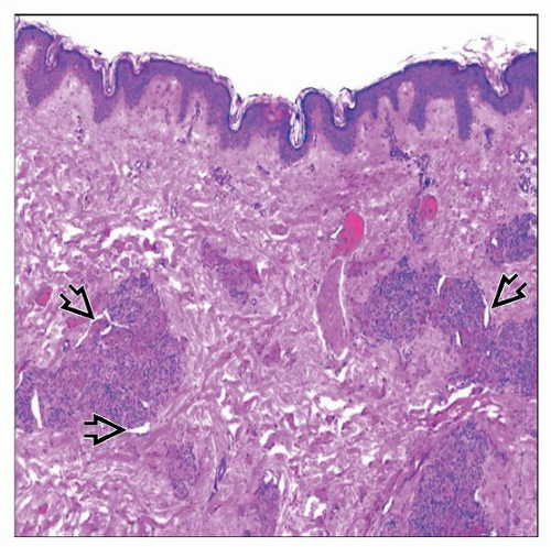 Scanning magnification of a tufted angioma shows scattered dermal lobular collections of vessels. The lobules bulge into dilated vessels, with a few peripheral semilunar spaces identified  . . |
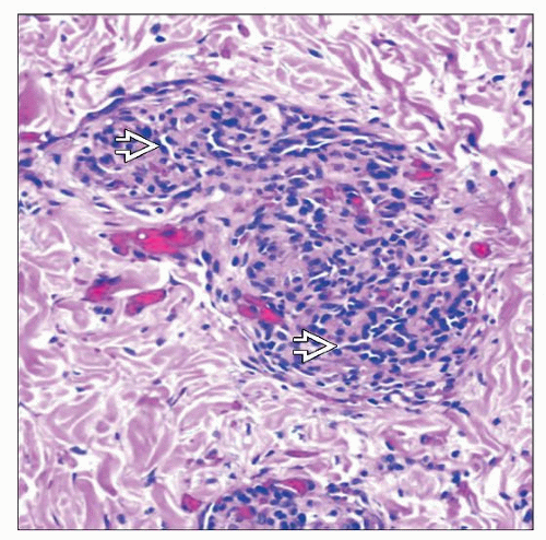 Higher magnification of a lobule shows a proliferation of small, slit-like vascular spaces  lined by bland oval to spindle-shaped endothelial cells. lined by bland oval to spindle-shaped endothelial cells. |
TERMINOLOGY
Synonyms
Acquired tufted angioma (ATA)
Angioblastoma (of Nakagawa)
Progressive capillary hemangioma
Tufted hemangioma
Definitions
Multiple cannonball-like, scattered cellular collections of small vessels in dermis
ETIOLOGY/PATHOGENESIS
Unknown
Most cases sporadic; rare familial case described
Some cases associated with pregnancy or liver transplantation


