Scanning magnification of acquired tufted angioma (ATA) shows scattered lobular collections of vessels  in the superficial and deep dermis. The lobules bulge into dilated vessels, with a few peripheral semilunar spaces identified.
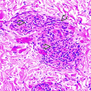
Higher magnification of ATA shows a lobular cluster of blood vessels lined by small, oval to spindle-shaped cells. Lumina are collapsed and slit-like
 .
.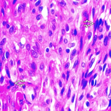
High-power view shows bland cytologic features of the tufted angioma endothelial cells. Some of the cells show mild nuclear hyperchromasia
 , but no significant cytologic atypia is identified.
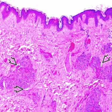
Scanning magnification of another ATA shows scattered dermal lobular collections of vessels. The lobules bulge into dilated vessels, with a few peripheral semilunar spaces identified
 .
.CLINICAL ISSUES
Epidemiology


Presentation


