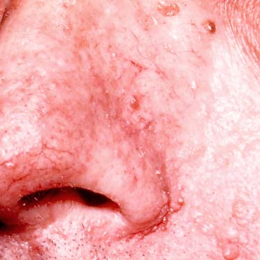
Clinical Photograph of Multiple Tricholemmomas Multiple tricholemmomas (TLs) are shown on and around the nose of a patient with Cowden syndrome. (Courtesy B. Hall, MD.)
Superficial Tricholemmoma This is a small, superficial tricholemmoma on the eyelid showing multifocal epidermal attachments . The tumor is composed of pale- to clear-staining epithelioid cells.
Superficial Tricholemmoma Higher magnification of a superficial TL shows a lobular proliferation of small, pale- to clear-staining adnexal cells. Note the thickened basement membrane surrounding the tumor .
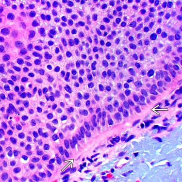
High Magnification of Tricholemmoma Basement Membrane High magnification of the periphery of a TL shows bland-appearing small cells with pale to clear-staining cytoplasm and small nucleoli. The peripheral cells show palisading, and there is a thickened peripheral basement membrane .


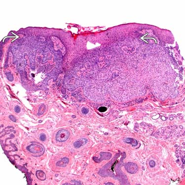
 . The tumor is composed of pale- to clear-staining epithelioid cells.
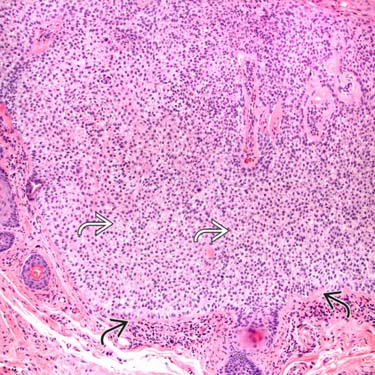
 adnexal cells. Note the thickened basement membrane surrounding the tumor
  .
.
 .
.


