• Rare recurrences and association with, or progression to, malignancy [basal cell carcinoma (BCC)]
Macroscopic
• Nodular lesion involving deep dermis and subcutis
• Large, typically > 1 cm in diameter
Microscopic
• Large, basaloid-appearing deep dermal-based nodule
Composed of irregular lobules and nests of basaloid cells
No epidermal connections
• Associated fibrotic stroma with increased numbers of fibroblasts
• Papillary mesenchymal bodies classically present, similar to TE
• Subtypes include trichogerminoma, rippled-pattern trichomatricoma, trichoblastic fibroma, and cutaneous lymphadenoma
Top Differential Diagnoses
• BCC
Usually shows multiple attachments to epidermis, mucinous stroma, and mitotic figures (focal or absent in TB)
• Trichoepithelioma
Smaller and more superficial than TB
• Sebaceoma
• Cylindroma
• Spiradenoma
Clinical Photograph of Trichoblastoma on Eyelid Clinical photograph shows a nodular lesion on the eyelid of a patient with a history of multiple trichoblastomas and trichoepitheliomas (TEs).
Scanning Magnification of Trichoblastoma Scanning magnification of a trichoblastoma shows a nodular basaloid proliferation associated with a fibrotic stroma. Note the presence of scattered folliculocystic structures .
High Magnification of Trichoblastoma High magnification of a trichoblastoma shows a lobular basaloid proliferation associated with a cellular, fibrotic stroma . Note the prominent stromal-stromal retraction artifact .
Trichoblastoma With Focal Sebaceous Differentiation Trichoblastoma can show sebaceous differentiation in some cases. Scattered clear cells with multivacuolated cytoplasm are identified .


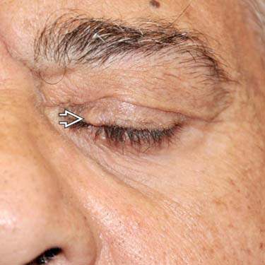
 of a patient with a history of multiple trichoblastomas and trichoepitheliomas (TEs).
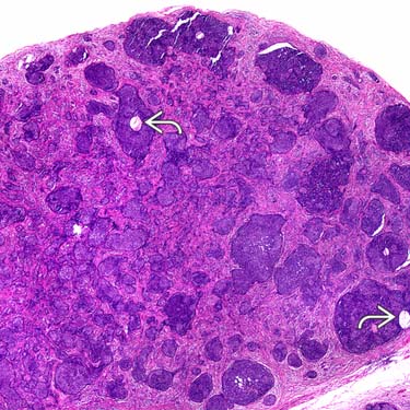
 .
.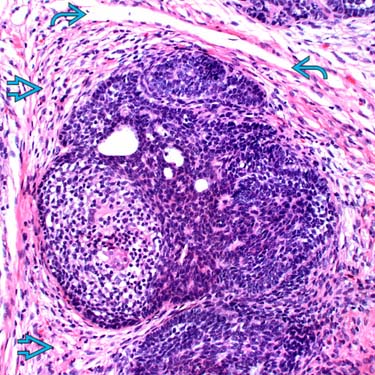
 . Note the prominent stromal-stromal retraction artifact
  .
.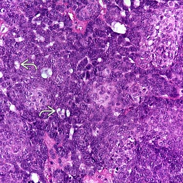
 .
.


