Trichoadenoma
Christine J. Ko, MD
Key Facts
Terminology
Benign follicular tumor
Clinical Issues
Often on the face
Solitary papule
Microscopic Pathology
Multiple mature cystic structures in dermis, showing epidermal-type keratinization
Cysts have granular layer and flaky central keratin
Interspersed among cystic structures are basaloid tubules and cords
Sclerotic stroma
Well circumscribed and not infiltrative
Top Differential Diagnoses
Trichoepithelioma
Trichofolliculoma
Microcystic adnexal carcinoma
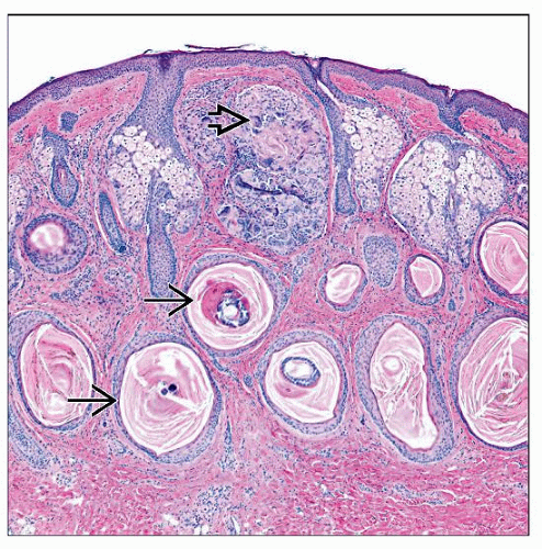 Low-magnification view of a trichoadenoma shows a dome-shaped lesion based in the dermis. Note numerous mature horn cysts  . There is focal granulomatous inflammation surrounding the keratin debris . There is focal granulomatous inflammation surrounding the keratin debris  . . |
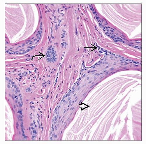 High magnification view of a trichoadenoma highlights the sclerotic stroma and the loose keratin debris within the horn cysts  . There are basaloid buds off of horn cysts . There are basaloid buds off of horn cysts  and in the stroma and in the stroma  . . |
TERMINOLOGY
Synonyms
Trichoadenoma of Nikolowski
Definitions
Benign follicular tumor
Predominance of mature folliculocystic structures in dermis
Epithelial tubules and cords interspersed
Sclerotic stroma
CLINICAL ISSUES


