Cellular Blue Nevi
Soheil Sam Dadras, MD, PhD
Olubukola Babalola
Key Facts
Terminology
Cellular variant of blue nevus
Clinical Issues
Occurs in children and young adults
Heavily pigmented black/blue nodule or plaque ranging from 1-2 cm; tends to occur on buttocks and sacrococcygeal region
Has low incidence of transformation into melanoma
Microscopic Pathology
Dumbbell-shaped architecture
Oval and spindle-shaped cells with bland cytology
Lack of mitoses or necrosis
Top Differential Diagnoses
Atypical cellular blue nevus
Melanoma developing in cellular blue nevus and malignant blue nevus
Desmoplastic melanoma
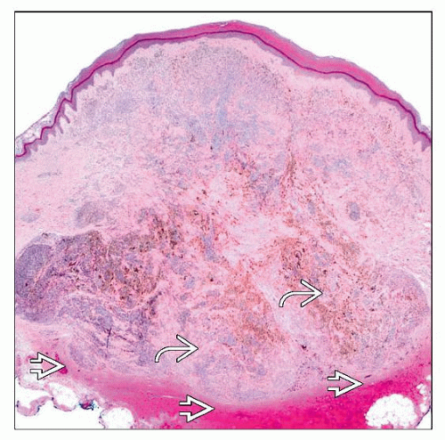 Scanning magnification of a CBN shows symmetrical hypercellular lobules resembling “dumbbells”  . These nodules push evenly and deeply beyond the reticular dermis into the subcutis . These nodules push evenly and deeply beyond the reticular dermis into the subcutis  . . |
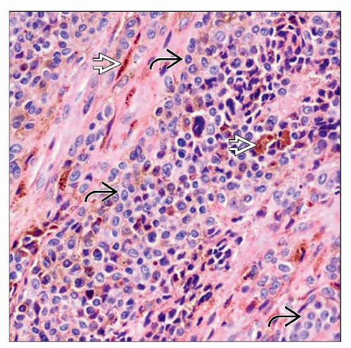 High-power examination of a hypercellular fascicle shows oval-to-spindled cells; when cut en face, these cells appear more cuboidal  . There are few scattered melanophages . There are few scattered melanophages  . . |
TERMINOLOGY
Abbreviations
Cellular blue nevus (CBN)
Definitions
Cellular variant of blue nevus
Stay updated, free articles. Join our Telegram channel

Full access? Get Clinical Tree


