Tinea Versicolor


Patient Story


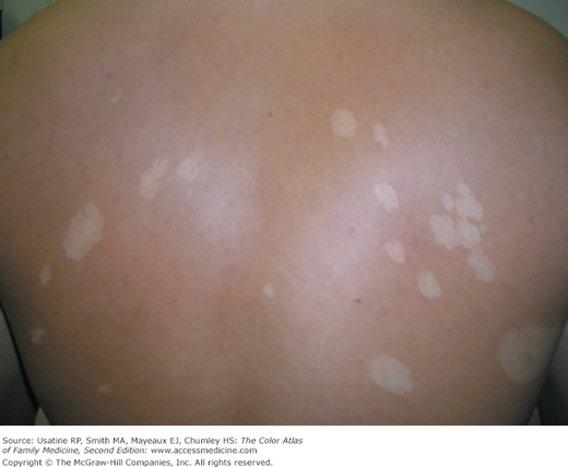
A young black man presents to the office with a 5-year history of white spots on his trunk (Figure 141-1). He denies any symptoms but worries if this could spread to his girlfriend. These spots get worse during the summer months but never go away completely. He was relieved to receive a treatment for his tinea versicolor and to find out that it is rarely spread to others through contact.


Figure 141-1


Tinea versicolor showing areas of hypopigmentation. (From the Usatine RP. What is in a name? West J Med. 2000;173(4):231-232.)


Introduction


Tinea versicolor is a common superficial skin infection caused by the dimorphic lipophilic yeast Pityrosporum (Malassezia furfur). The most typical presentation is a set of hypopigmented macules and patches with fine scale over the trunk in a cape-like distribution.


Synonyms


Pityriasis versicolor is actually a more accurate name as “tinea” implies a dermatophyte infection. Tinea versicolor is caused by Pityrosporum and not a dermatophyte.


Epidemiology


  • Seen more commonly in men than in women.
  • Seen more often during the summer, and is especially common in warm and humid climates.


Etiology and Pathophysiology


  • Tinea versicolor is caused by Pityrosporum (M. furfur), which is a lipophilic yeast that can be normal human cutaneous flora.
  • Pityrosporum exists in two shapes—Pityrosporum ovale (oval) and Pityrosporum orbiculare (round).
  • Tinea versicolor starts when the yeast that normally colonizes the skin changes from the round form to the pathologic mycelial form and then invades the stratum corneum.1
  • Pityrosporum is also associated with seborrhea and Pityrosporum folliculitis.
  • The white and brown colors are secondary to damage caused by the Pityrosporum to the melanocytes, while the pink is an inflammatory reaction to the organism.
  • Pityrosporum thrive on sebum and moisture; they tend to grow on the skin in areas where there are sebaceous follicles secreting sebum.


Diagnosis


Clinical Features


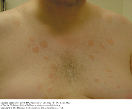
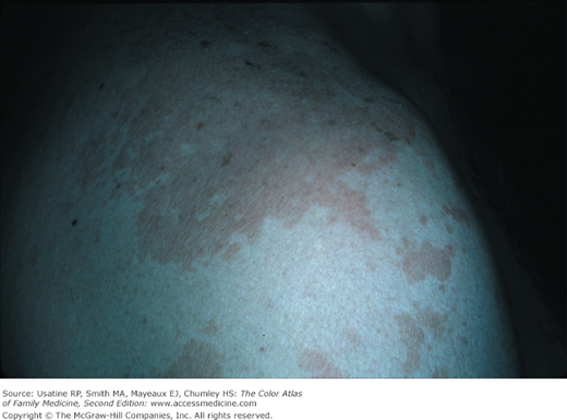
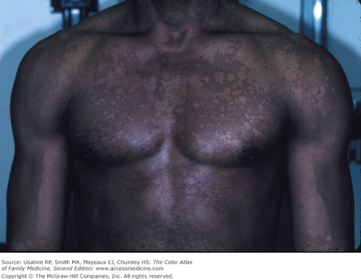
Tinea versicolor consists of hypopigmented, hyperpigmented, or pink macules and patches on the trunk that are finely scaling and well-demarcated. Versicolor means a variety of or variation in colors; tinea versicolor tends to come in white, pink, and brown colors (Figures 141-1, 141-2, 141-3, 141-4, 141-5).


Figure 141-2


Patches of hypopigmentation across the back caused by tinea versicolor in a young Latino man. Vitiligo is on the differential diagnosis in this case. A KOH preparation confirmed tinea versicolor. (Courtesy of Richard P. Usatine, MD.)


Figure 141-3


Pink scaly patches caused by tinea versicolor. Seborrhea may be seen in this location, but tends to be worse in the presternal region. (Courtesy of Richard P. Usatine, MD.)


Figure 141-4


Large areas of pink tinea versicolor on the shoulder in a cape-like distribution. (Courtesy of Richard P. Usatine, MD.)


Jun 5, 2016 | Posted by in GENERAL & FAMILY MEDICINE | Comments Off on Tinea Versicolor

Full access? Get Clinical Tree

Get Clinical Tree app for offline access