Upon completion of this chapter, the student should be able to do the following: • Describe the general structure of vertebrae and identify the structural components on a diagram. • State the characteristics that distinguish cervical vertebrae from other types. • Distinguish the first two cervical vertebrae from other cervical vertebrae and state their specific names. • Identify the characteristic features that distinguish thoracic vertebrae from other vertebrae. • Identify the characteristic features that distinguish lumbar vertebrae from other vertebrae. • Describe the structural features of the sacrum and coccyx. • Discuss the composition and purpose of intervertebral discs. • Identify the curvatures of the vertebral column and state the direction of the curvatures. • Identify three types of abnormal curvatures of the vertebral column. • Describe and state the location of the following ligaments associated with the vertebral column: anterior and posterior longitudinal ligaments, ligamenta flava, interspinous ligaments, supraspinous ligaments, ligamentum nuchae, and intertransverse ligaments. • Identify the muscles in the superficial, intermediate, and deep layers of the intrinsic back muscles. • Describe the meninges of the spinal cord. • Identify the structural features of the spinal cord in longitudinal and cross-sectional views. • Name the five groups of spinal nerves and state the number of nerves in each group. • Distinguish between dorsal and ventral nerve roots and state the components of each. • Describe the location, components, and functions of the four major nerve plexuses and name the principal nerves that emerge from each plexus. • Describe the vasculature of the spinal cord. • Identify the margins of the anterior and posterior triangles of the neck and name the principal components in each triangle. • Identify the regions of the pharynx by describing the location and features of each region. • Describe the features of the larynx. • Discuss the relationships of the esophagus and trachea as they descend through the neck. • Describe the location of the thyroid and parathyroid glands relative to the trachea. • Discuss the relationships of the internal jugular vein to other vessels and anatomical structures as it descends from the jugular foramen to the brachiocephalic vein. • Describe the pathways and relationships of the common carotid, external carotid, and internal carotid arteries. • State the origin and pathway of the vertebral arteries. • Explain what is meant by the term sympathetic trunk and describe its location. • Name one cranial nerve located in the neck. • Identify the nerve plexus located in the neck and name one nerve that emerges from this plexus. • Describe the composition and location of the brachial plexus and name the region innervated by the nerves that emerge from this plexus. • Identify the features of the neck, including vertebrae, muscles, viscera, and blood vessels, in transverse and midsagittal sections. Key Terms, Structures, and Features to Be Identified and/or Described Anterior and posterior longitudinal ligaments Columns of white matter in spinal cord Dorsal (posterior) median sulcus Horns of gray matter in spinal cord Levator costarum muscle Nerve plexus Posterior scalene muscle Scapula Superior and inferior articular processes Ventral (anterior) median fissure All vertebrae have a common structural pattern (Fig. 6-1), although variations exist between them. The thick, anterior, weight-bearing portion of a vertebra is the body or centrum The posterior, curved portion is the vertebral arch Together, the body and vertebral arch surround a central opening, the vertebral foramen When the vertebrae are stacked together, the vertebral foramina make a vertebral canal, which contains and protects the spinal cord. The vertebral arch is formed by the transverse processes, the spinous process, the pedicles, the laminae, and the superior and inferior articular processes. Transverse processes project laterally from the vertebral arch, and the spinous process is in the posterior midline. These processes are places for muscle attachment. The spinous processes can be felt as bony projections along the midline of the back. The portion of the vertebral arch adjacent to the body, between the body and the transverse process, is the pedicle A concave surface on the upper and lower margins of the pedicles is called the vertebral notch When the superior and inferior vertebral notches of adjacent vertebrae meet, they form intervertebral foramina, which transmit spinal nerves and blood vessels. The portion between the transverse process and the spinous process is the lamina Superior and inferior articular processes project superiorly and inferiorly, respectively, from the vertebral arch. The superior articular process of one vertebra articulates with the inferior articular process of the preceding vertebra in the column. Fig. 6-2 is a radiograph of cervical vertebrae that demonstrates some of these features. The seven cervical vertebrae are designated C1 through C7. In general, the cervical vertebrae (Figs. 6-3 and 6-4) can be distinguished from other vertebrae because the cervicals have transverse foramina in the transverse processes. The foramina allow passage of the vertebral arteries as they ascend through the neck to the brain. Also, the spinous processes of cervical vertebrae are forked, or bifid. The exception to this is the seventh cervical vertebra (C7), which has a long spinous process that typically is not bifid. The C7 spinous process is easily palpable at the base of the neck. The first two cervical vertebrae are modified and have no disc between them. The atlas (C1), shown in Figs. 6-5 and 6-6, has no body and no spinous process, and it has short transverse processes. It is a ring that consists of an anterior arch, a posterior arch, and two large lateral masses that have large articular facets. The superior facets articulate with the occipital condyles on the occipital bone, and the inferior facets articulate with the vertebra below. The axis (C2), shown in Figs. 6-7 to 6-9, has a dens, or odontoid process, that projects upward from the vertebral body like a tooth. The odontoid process acts as a pivot for rotation of the atlas. The 12 thoracic vertebrae are designated T1 through T12. These can be distinguished from other vertebrae by the costal facets, located on the bodies and transverse processes, for articulation with the ribs. The head of the rib articulates with the vertebral body, and the tubercle of the rib articulates with the transverse process. They also have long, pointed spinous processes. These features are illustrated in Fig. 6-10. The axial CT image in Fig. 6-11 shows the articulations of a thoracic vertebra with ribs. The sagittal CT image in Fig. 6-12 shows the long, pointed spinous processes that are typical of thoracic vertebrae. The five lumbar vertebrae, designated L1 through L5, make up the part of the vertebral column in the small of the back. The lumbar vertebrae (Figs. 6-13 and 6-14) have large, heavy bodies because they support most of the body weight and have many back muscles attached to them. They also have short, blunt spinous processes. The sagittal MR image in Fig. 6-15 shows the thick, heavy bodies of the lumbar vertebrae and the intervertebral discs between the vertebrae. The sacral region of the vertebral column (Fig. 6-16) consists of five vertebrae that fuse to form the sacrum The transverse processes of the vertebrae fuse to form the lateral masses (alae) which articulate with the pelvic girdle laterally at the sacroiliac joints Within the lateral masses, sacral foramina allow for passage of nerves. The first sacral segment has a prominent ridge, called the sacral promontory, on the visceral surface. The region inferior to this landmark is the true pelvic cavity. The spinous process of the S5 segment is absent, leaving an opening called the sacral hiatus The coronal CT image in Fig. 6-17 shows these features. The coccyx, or tailbone, is the most inferior region of the vertebral column. The child has four separate small bones (the number can vary from three to five), which fuse to form a single bone in the adult. Several muscles have some point of attachment on the coccyx. The CT reconstruction in Fig. 6-18 shows features of the sacrum and coccyx. The intervertebral discs, located between the vertebral bodies, are fibrocartilaginous pads classified as symphysis joints; they are designed for strength. In addition to the discs, the vertebral bodies are joined by ligaments. Each disc consists of a soft central core, called the nucleus pulposus, and a firm outer ring, called the anulus fibrosus Fig. 6-19 illustrates an intervertebral disc. The sagittal MR image in Fig. 6-20 shows lumbar vertebrae with intervertebral discs. The nucleus pulposus acts as a shock absorber and as a ball bearing during flexion, extension, and lateral bending of the vertebral column. The anulus fibrosus is composed of rings of fibrocartilage that run obliquely from one vertebra to another to form strong bonds between the vertebrae. The intervertebral discs are thickest in the cervical and lumbar regions, which provides greater flexibility in these regions. As people get older, the nucleus pulposus may become thinner. This accounts for some of the loss of height that occurs as a result of aging. Changes also may occur in the anulus fibrosus that permit the nucleus pulposus to protrude through the outer ring; this is called a herniated disc (Fig. 6-21). The protrusion may compress an adjacent spinal nerve and cause pain in the lower back and/or leg. Normally, when viewed from the side (Fig. 6-22), the vertebral column has four curvatures. These curvatures increase the strength, resilience, and flexibility of the vertebral column. The thoracic curvature and the sacral curvature are concave anteriorly (convex posteriorly) and are present at birth. The cervical and lumbar curvatures are convex anteriorly (concave posteriorly). The cervical curvature develops when the baby holds its head erect, and the lumbar curvature develops when the child begins to stand. Fig. 6-23 is a radiograph of the lumbar spine that shows the anteriorly convex curvature. Several types of abnormal variations occur in the curvatures of the vertebral column. Some of these are congenital, whereas others may be due to poor posture, disease, or unequal muscle pull on the column. Scoliosis is an abnormal lateral curvature that most commonly occurs in the thoracic region. If left untreated, it may become severe and result in breathing difficulties. Kyphosis is an exaggerated dorsal curvature in the thoracic region. It is frequently seen in older individuals as a result of osteoporosis. It also may be due to a bone deformity known as rickets. Lordosis is an exaggerated lumbar curvature. This may occur temporarily in pregnant women when they throw back their shoulders and accentuate the lumbar curvature in an attempt to maintain their center of gravity. Fig. 6-24 illustrates abnormal spinal curvatures. The vertebral column is held in place by straplike ligaments. The major supporting ligaments are the anterior and posterior longitudinal ligaments The anterior longitudinal ligament begins at C1 and extends downward along the entire anterior surface of the vertebral bodies to the sacrum. It is strongly attached to the bony vertebral bodies and to the discs to maintain stability of the joints and to prevent hyperextension (bending too far backward). The posterior longitudinal ligament is a narrow ligamentous band that runs inside the vertebral canal, along the posterior surface of the vertebral bodies, for the entire length of the vertebral column. It is somewhat weaker than the anterior longitudinal ligament. The ligament is attached to the intervertebral discs and the posterior edges of the vertebral bodies. The posterior longitudinal ligament tends to prevent hyperflexion (bending too far forward) of the vertebral column. Ligamenta flava are short bands of yellow elastic fibers on each side of the spinous process. These ligaments connect the laminae of adjacent vertebrae and help to maintain the normal curvature of the vertebral column. The margins of adjacent spinous processes from C7 to the sacrum are joined by interspinous ligaments. Supraspinous ligaments join the tips of the spinous processes in the same region. From C7 upward to the occipital bone, the interspinous and supraspinous ligaments are represented by the ligamentum nuchae Intertransverse ligaments connect adjacent transverse processes. These consist of a few scattered ligamentous fibers, except in the lumbar region, where they are membranous. The ligaments associated with the vertebral column are summarized in Table 6-1. Fig. 6-25 illustrates some of the ligaments. TABLE 6-1 Ligaments of the Vertebral Column The muscles associated with the back are arranged in three layers. The superficial and intermediate layers are extrinsic muscles and are concerned with respiration and movement of the limbs. These muscles, described in other portions of this book, include the trapezius, the latissimus dorsi, the levator scapulae, the rhomboids, and the serratus posterior. The deep muscle layer includes the true back muscles; these are the intrinsic muscles, which are concerned with the maintenance of posture and movements of the vertebral column. The intrinsic muscles are summarized in Table 6-2. TABLE 6-2 Intrinsic Muscles Associated With the Vertebral Column
The Vertebral Column, Spinal Cord, and Neck
Anatomical Review of the Vertebral Column
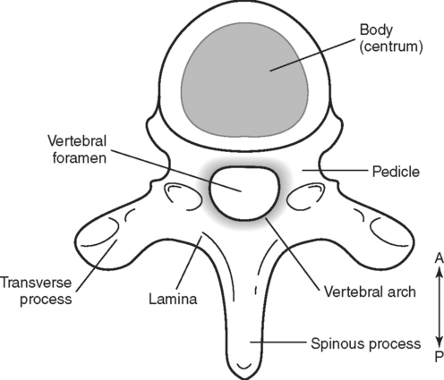
Structure of the Vertebrae
General Structure
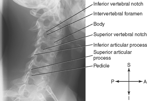
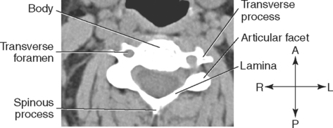
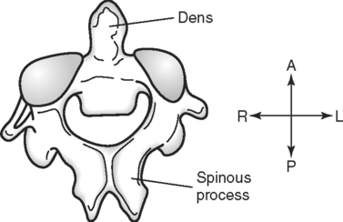
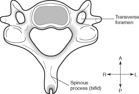
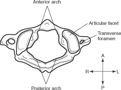
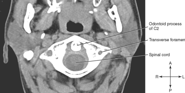
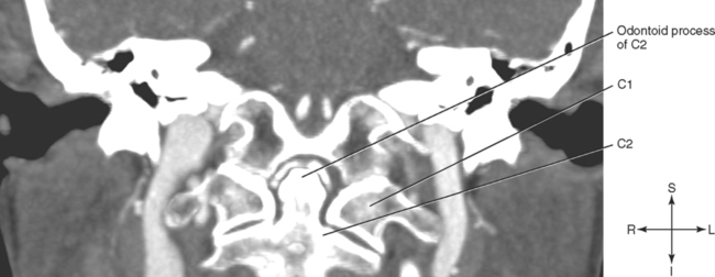
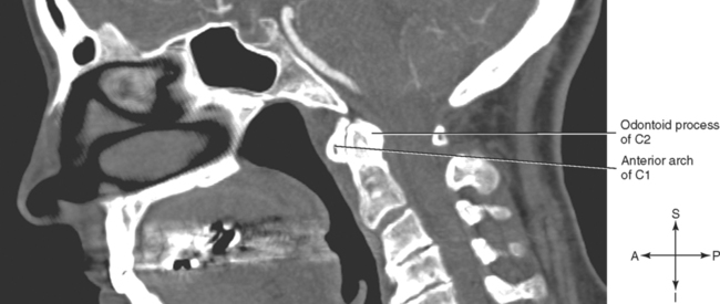
Cervical Vertebrae
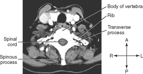
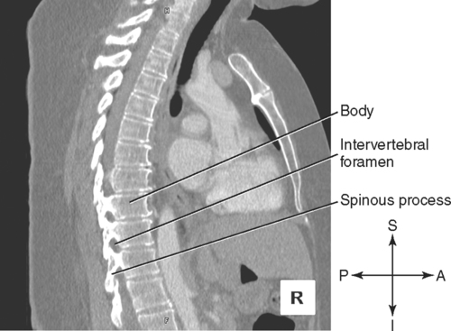
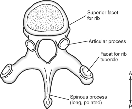
Thoracic Vertebrae
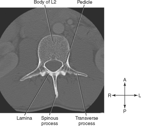
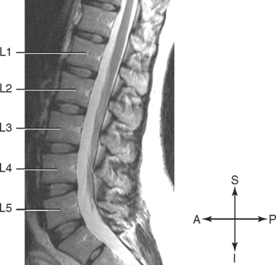
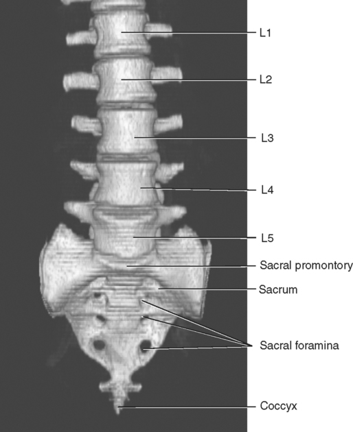
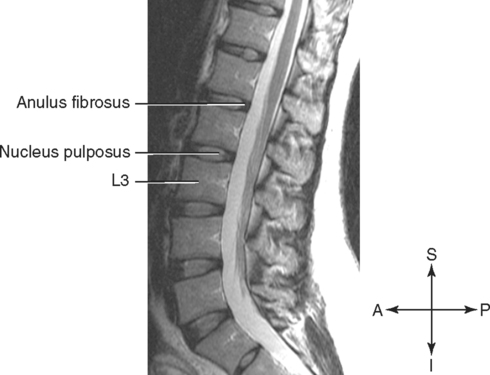
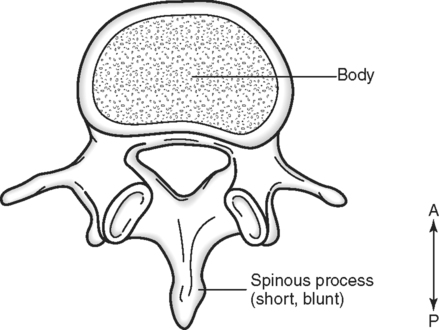
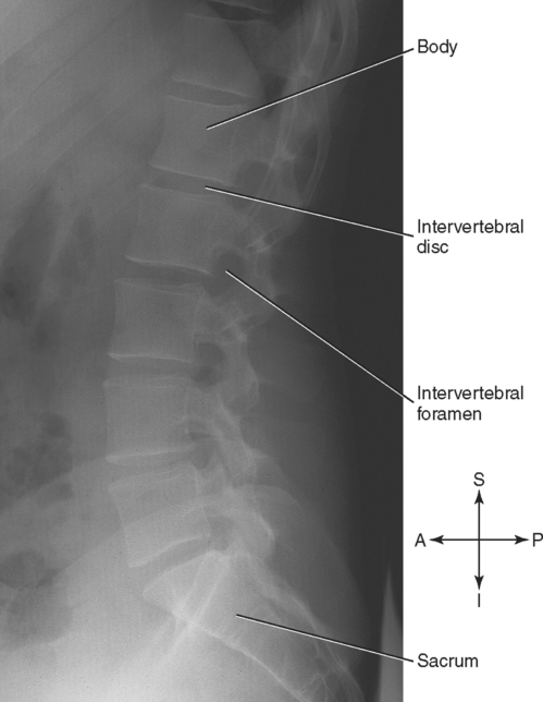
Lumbar Vertebrae
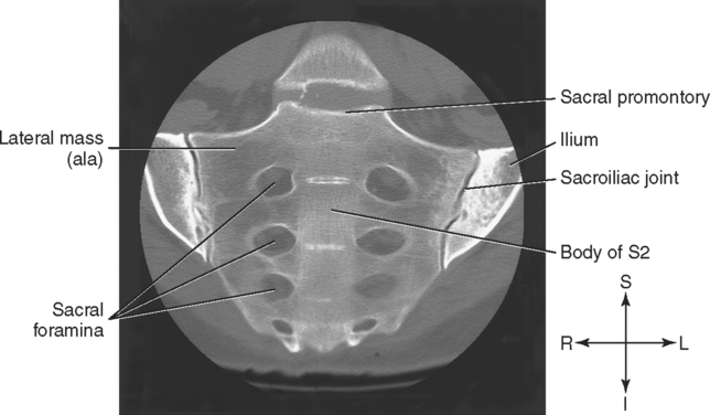
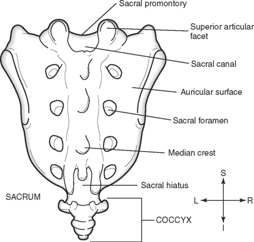
Sacrum and Coccyx
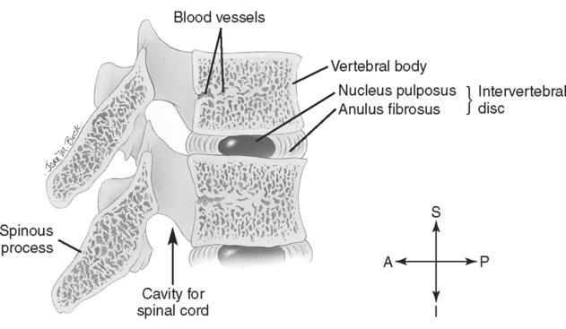
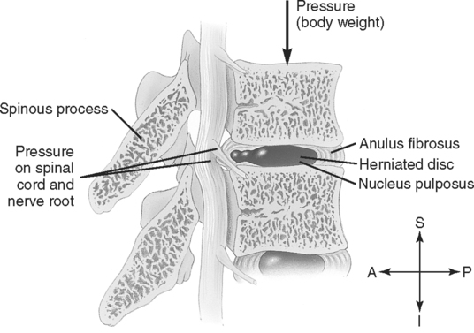
Intervertebral Discs
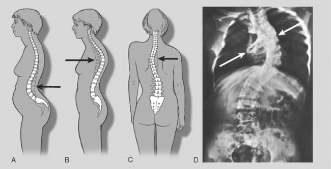
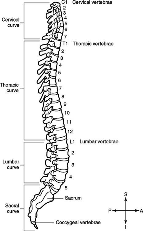
Curvatures of the Vertebral Column
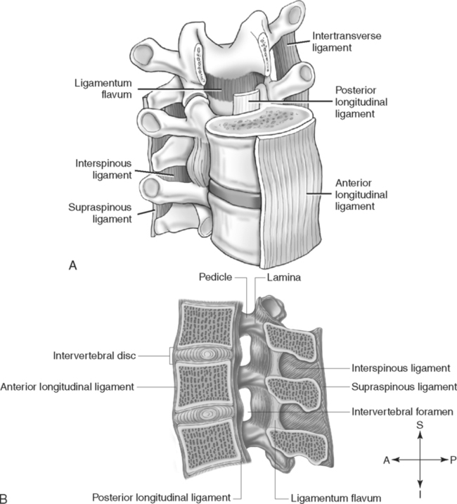
Ligaments of the Vertebral Column
Ligament
Description and Location
Anterior longitudinal ligament
Extends from C1 to sacrum; attached to anterior surface of vertebral bodies and discs; prevents hyperextension
Posterior longitudinal ligament
Extends from C1 to sacrum; narrow band that is attached to posterior edges of vertebral bodies and discs; located within vertebral canal; prevents hyperflexion
Ligamenta flava
Short bands that connect laminae of adjacent vertebrae on each side of spinous process; help maintain normal curvature of column
Interspinous ligaments
Short bands that connect the margins of adjacent spinous processes in thoracic and lumbar regions
Supraspinous ligaments
Short bands that connect the tips of adjacent spinous processes in thoracic and lumbar regions
Intertransverse ligaments
Short bands that connect adjacent transverse processes; poorly developed except in lumbar region
Ligamentum nuchae
Combination of interspinous and supraspinous ligaments in the cervical region
Muscles Associated With the Vertebral Column
Muscle
Origin
Insertion
Function
Description
Superficial Layer
Splenius capitis
Ligamentum nuchae and spinous processes of C7 to T6
Mastoid process of temporal bone and adjacent occipital bone
Extend the head; rotate neck if one side contracts
Superficial intrinsic muscle in the neck
Splenius cervicis
Ligamentum nuchae and spinous processes of C7 to T6
Transverse processes of C2 to C4
Extend the head; rotate neck if one side contracts
Superficial intrinsic muscle in the neck
Intermediate Layer
Iliocostalis
Iliac crests and ribs
Angles of ribs and transverse processes of C4 to C6
Extend vertebral column to maintain posture; if one side contracts, column bends to that side
Most lateral column of the erector spinae muscle group
Longissimus
Transverse processes of lumbar through cervical vertebrae
Transverse processes of thoracic and cervical vertebrae, ribs superior to origin, and mastoid process of temporal bone
Extend vertebral column; if one side contracts, column bends to that side
Intermediate column of erector spinae muscle group
Spinalis
Spinous processes of upper lumbar and lower thoracic vertebrae
Spinous processes of upper thoracic and cervical vertebrae
Extend vertebral column
Most medial column of erector spinae muscle group
Deep Layer
Semispinalis
Transverse processes of C7 to T12
Occipital bone, spinous processes of cervical and thoracic vertebrae
Extend vertebral column
Composite muscle of deep layer; extends from thoracic region to head; part of the transversospinal group
Multifidus
Transverse processes of vertebrae
Spinous processes of preceding vertebrae
Stabilize vertebral column and rotate to opposite side
Short muscle bundles that pass superiorly over two to five vertebrae and then insert; part of the transversospinal group
Rotatores
Transverse processes of vertebrae
Spinous process of vertebra immediately superior to origin
![]()
Stay updated, free articles. Join our Telegram channel

Full access? Get Clinical Tree

 Get Clinical Tree app for offline access