Introduction to Sectional Anatomy
Upon completion of this chapter, the student should be able to do the following:
• Identify the transverse, sagittal, midsagittal, parasagittal, and frontal planes.
• Describe the anatomical position.
• Use correct directional terms to describe the relative position of one body part to another.
• Describe the location, subdivisions, and contents of the dorsal body cavity and the ventral body cavity.
• Name and identify the nine abdominopelvic regions.
• Distinguish between connective tissue membranes and epithelial membranes and give two examples of each type.
• Describe the location of mucous membranes and serous membranes.
• Distinguish between the visceral and parietal layers of serous membranes.
• State the specific locations of the parietal and visceral layers of the pleura, pericardium, and peritoneum.
• Use correct regional terms to refer to specific areas of the body.
Key Terms, Structures, and Features to Be Identified and/or Described
Antebrachial region
Axial plane
Axillary region
Buccal region
Cephalic region
Costal region
Cranial region
Cubital region
Femoral region
Gluteal region
Occipital region
Oral region
Pectoral region
Pelvic region
Pericardium
Popliteal region
Sternal region
Thigh
Thoracic region
Vertebral region
Comparison of Traditional Anatomy and Sectional Anatomy
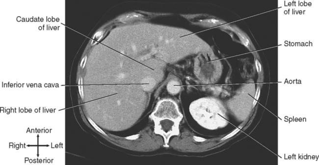
For orientation of a transverse (axial) slice, when viewing either the inferior of a cadaveric slice or a real image, imagine that the patient is supine and you are standing at the person’s feet looking up into the patient’s body. This means that your right is the patient’s left. Fig. 1-1 shows a transverse CT image through the abdomen that illustrates this orientation. The right lobe of the liver, inferior vena cava, and caudate lobe of the liver are on the patient’s right side, but they are on the left side of the image. The left lobe of the liver, the stomach, the spleen, and the left kidney are on the patient’s left side, but they are on the right side of the image. The aorta is slightly to the left of midline. Correct orientation is critical to proper identification of structures.
Now, a couple of cautionary notes for learning sectional anatomy:
1. A given organ varies in appearance at different levels in the same individual. Fig. 1-2 shows images of the liver at three different levels. Notice the changes in appearance at the different levels.
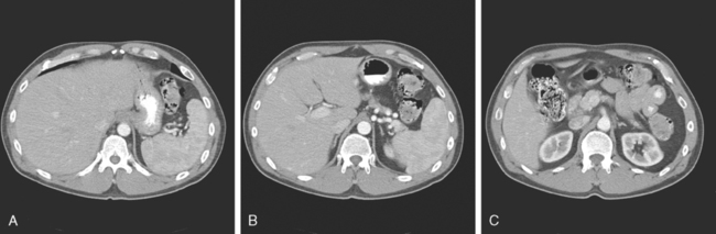
2. Attempting to memorize images likely will lead to mistakes, because no two images are the same. Every individual is different, and even images of the same individual taken at the same level differ because of breathing and involuntary movements. The key is understanding, not memorization.
Body Planes
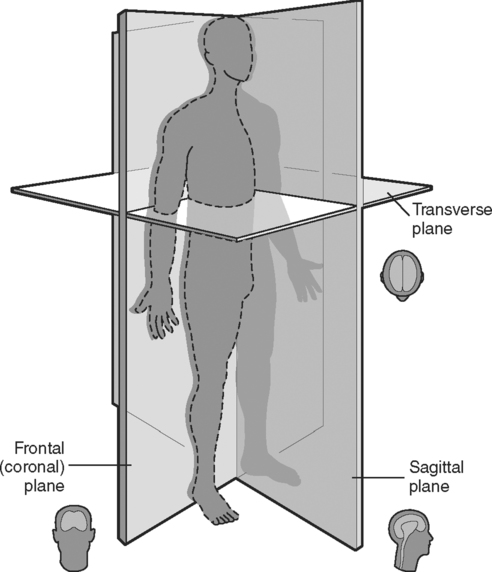
To visualize the spatial relationships of internal body parts, the body is sectioned, or cut, along a flat surface, or plane. The ability to interpret sections in various planes is becoming increasingly important in the clinical sciences, especially in the imaging sciences. The three most commonly used planes are the transverse (axial), sagittal, and frontal (coronal) planes, which are at right angles to each other (Fig. 1-3). By analyzing body parts in all three planes, you develop an understanding of the anatomical relationships within the body.
Stay updated, free articles. Join our Telegram channel

Full access? Get Clinical Tree


